
This Key Event Relationship is licensed under the Creative Commons BY-SA license. This license allows reusers to distribute, remix, adapt, and build upon the material in any medium or format, so long as attribution is given to the creator. The license allows for commercial use. If you remix, adapt, or build upon the material, you must license the modified material under identical terms.
Relationship: 3487
Title
Decrease, intratesticular testosterone leads to nipple retention, increased
Upstream event
Downstream event
Key Event Relationship Overview
AOPs Referencing Relationship
| AOP Name | Adjacency | Weight of Evidence | Quantitative Understanding | Point of Contact | Author Status | OECD Status |
|---|---|---|---|---|---|---|
| Decreased testosterone synthesis leading to increased nipple retention (NR) in male (rodent) offspring | non-adjacent | High | Terje Svingen (send email) | Under development: Not open for comment. Do not cite |
Taxonomic Applicability
Sex Applicability
| Sex | Evidence |
|---|---|
| Male | High |
Life Stage Applicability
| Term | Evidence |
|---|---|
| Foetal | High |
Key Event Relationship Description
This non-adjacent KER describes a fetal decrease in intratesticular testosterone leading to NR in male offspring. In this KER, intratesticular testosterone includes measurements of testosterone in homogenates of testes after in vivo exposure to chemicals, as well as measurements of testosterone production in testes ex vivo from exposed animals.
In male mammals, the testes are the first sex organs to develop. Once formed, they produce testosterone by steroidogenesis. The adrenal glands have been shown to synthesize testosterone, but on a much smaller scale, and the testes are the main site of testosterone production (Naamneh Elzenaty et al., 2022). Testicular testosterone is secreted into the blood to initiate masculinization of the peripheral reproductive tissues. In rats and mice, this includes effects on the developing mammary glands, which develop sexually dimorphic. Testosterone either directly activates the AR in the mammary glands or is converted to the more potent androgen dihydrotestosterone (DHT) (Murashima et al., 2015). Activation of AR by androgens in the mammary glands causes apoptosis of epithelial cells and thus separation of the glands from the overlying epidermis. Consequently, no nipples are formed (Kratochwil, 1986). During low androgen levels, such as in female rodents, nipple development progresses to form up to 10 (mice) and 12 (rats) nipples.
As suppression of nipple development in male rats and mice is dependent on androgens, marked reductions in testicular testosterone production can thus cause nipple retention.
The KER is not directly applicable to humans, as both males and females have two nipples, and there is no known effect of androgens on their development (Schwartz et al., 2021). However, NR is a clear readout of reduced androgen action and fetal masculinization during development, which is relevant to humans (Schwartz et al., 2021). It is included as a mandatory endpoint in several rodent OECD Test Guidelines (OECD, 2025a, 2025b, 2025c) and considered an adverse outcome applicable to the setting of Points of Departure for use in human health risk assessment (OECD, 2013).
Evidence Collection Strategy
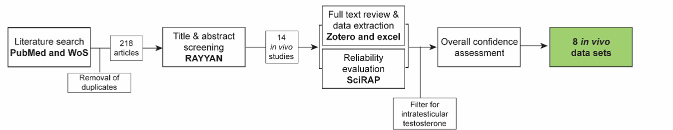
A systematic approach (Fig. 1,60mdu2onwj_KER3487_Figure_1.png (1347×261)) was used to collect evidence based on the methodology described in (Holmer et al., 2024). The evidence collection for this KER was done concurrently with the evidence collection for KER-3486 ‘decreased circulating testosterone leads to increased nipple retention’, for which the same search string was used.
Search strategy
Search strings were synthesized for PubMed and Web of Science Core Collection based on the review question, ‘Does decreased testosterone during fetal development lead to increased nipple retention in male mammals?’
Search string in PubMed: "testosterone*" AND ("nipple*” OR “areola*”)
Search string in Web of Science: "testosterone*" AND ("nipple*” OR “areola*”)
The searches were performed on 01.10.2024
Title & abstract screening:
Retrieved articles were screened in the online tool RAYYAN https://www.rayyan.ai/. After removal of duplicates, the titles and abstracts of the remaining 218 articles were screened according to pre-defined inclusion and exclusion criteria:
Inclusion criteria:
- In vivo studies in male mammals where fetal testosterone is reduced and nipples/areolas are measured *
- Reviews on nipple/areola retention
- In vitro, ex vivo, and in vivo mechanistic studies on nipple/areola retention
Exclusion criteria:
- Papers not in English
- Abstracts and other non-full text publications
*In cases where this criterion could not be determined by reading the abstract, the study was included for full text review
Full text review, data extraction and reliability evaluation of animal studies:
For the in vivo studies, the full text papers were reviewed using the same exclusion criteria as in the title & abstract screening, and data were extracted from the included papers into an Excel template. In parallel, methodological reliability was assessed using the online tool Science in Risk Assessment and Policy (SciRAP; http://www.scirap.org, see appendix 1, 2p6ci7kafm_KER_3487_Appendix_1.pdf). Based on the SciRAP evaluations, animal studies were assigned a reliability category using the principles outlined in table 1. Studies were divided into different datasets if multiple different chemicals, different exposure windows, or different timepoints of measurement of NR were included.
Moreover, as this KER was made in parallel with several other KERs for other male reproductive endpoints (anogenital distance and hypospadias), five studies retrieved in the searches for these KERs, which also measured NR, but were not detected in the search for this KER were also added, data extracted and evaluated for reliability.
The collected data were then filtered to only include data sets measuring intratesticular testosterone, either in whole testis or production during ex vivo testis culture. Mixture studies were excluded from the final evidence as the chemicals may have many different upstream mechanisms.
The overall strength of the empirical support was assessed according to the principles outlined in the AOP wiki handbook. Only studies in reliability categories 1 (reliable without restriction) and 2 (reliable with restriction) were used for the assessment of overall confidence in the data.
Table 1 Principles for translation SciRAP evaluations into reliability categories.
|
Reliability Category |
Principles for Categorization |
|
1.Reliable without restriction |
SciRAP methodological quality Score > 80 and all key criteria1 are “Fulfilled” and there are no deficiencies in the non-key criteria that might affect study reliability. |
|
2. Reliable with restriction |
SciRAP methodological quality Score > 65 and one or several of the key criteria are “Partially Fulfilled” or there are minor deficiencies in the non-key criteria that might affect study reliability. |
|
3. Not reliable |
SciRAP methodological quality Score < 65 or one or several of the key criteria are “Not Fulfilled” or there are major deficiencies in the non-key criteria that affect study reliability. |
|
4. Not assignable |
Two or more of the key criteria are “Not Determined” |
1Key criteria were SciRAP criteria for methodological quality judged as specifically critical for the reliability of the data for this KER and were determined a priori. The key criteria for this data collection are outlined in Appendix 1, 2p6ci7kafm_KER_3487_Appendix_1.pdf.
Evidence Supporting this KER
Biological Plausibility
The biological plausibility for this KER is judged to be high given the canonical biological knowledge on normal reproductive development in rodents.
In common strains of rats and mice, females have 12 and 10 pairs of nipples, respectively, while males usually do not have nipples, although in rare instances control male rats may display a retained nipple (Schwartz et al., 2021). The sexual dimorphism of the mammary tissue is regulated by the androgens testosterone and DHT, during fetal life. Testosterone is produced by the fetal testes by the steroidogenesis pathway, starting from ~GD15. The testes are the primary male sex organs and produce most of the circulating testosterone, although a minor part may be contributed by other organs such as the adrenals (Naamneh Elzenaty et al., 2022). DHT is produced from testosterone in peripheral tissues by the enzyme 5α-reductase. Both testosterone and DHT activate AR in reproductive tissues to initiate masculinisation. The programming of the tissues mainly happens within the masculinization programming window (GD16-20 in rats) (Murashima et al., 2015; Welsh et al., 2014).
The mammary glands start out the same in both sexes, developing along two milk lines in early fetal life. In males, androgens activate AR in the mammary gland mesenchymal cells, and in turn, the cells activate apoptosis of epithelial cells that otherwise would contribute to the development of nipples (Kratochwil, 1986; Schwartz et al., 2021).
Given the dependency of testosterone for regression of the nipples, either through direct AR activation or conversion to DHT, it is highly plausible that a decrease in intratesticular testosterone levels will lead to nipple retention in males.
Empirical Evidence
The empirical evidence from studies in animals for this KER is overall judged as strong.
From the data collection, 8 data sets were extracted. The data sets included different stressors causing reduced fetal intratesticular testosterone, all in rats (Table 2 and Appendix 2, 91ldxzzu4f_KER_3487_Appendix_2_clean.pdf). Of these 8 data sets, seven showed concurrent nipple retention.
Table 2 Empirical evidence for KER 3487 LOAEL: Lowest observed adverse effect level; NOAEL: No observed adverse effect level. See Appendix 2, for specifications.
|
Species |
Stressors(s) |
Effect on upstream event (circulating testosterone) |
Effect on downstream event (NR) |
Reference |
|
Rat |
Butyl benzyl phthalate |
LOAEL 500 mg/kg bw/day |
No effect NOAEL 500 mg/kg bw/day |
(Hotchkiss et al., 2004) |
|
Rat |
Dibutyl phthalate |
LOAEL 500 mg/kg bw/day |
LOAEL 500 mg/kg bw/day |
(Martino-Andrade et al., 2009) |
|
Rat |
Diethylhexyl phthalate |
LOAEL 750 mg/kg bw/day |
LOAEL 750 mg/kg bw/day |
(Borch et al., 2004) |
|
Rat |
Diisonyl phthalate |
LOAEL 600 mg/kg bw/day |
LOAEL 750 mg/kg bw/day |
(Boberg et al., 2011) |
|
Rat |
Linuron |
LOAEL 75 mg/kg bw/day |
LOAEL 75 mg/kg bw/day |
(Hotchkiss et al., 2004) |
|
Rat |
Prochloraz |
LOAEL 50 mg/kg bw/day |
LOAEL 50 mg/kg bw/day |
(Laier et al., 2006) |
|
Rat |
Prochloraz |
LOAEL 30 mg/kg bw/day |
LOAEL 30 mg/kg bw/day |
(Vinggaard et al., 2005) |
|
Rat |
Tebuconazole |
LOAEL 100 mg/kg bw/day |
LOAEL 50 mg/kg bw/day |
(Taxvig et al., 2007) |
Dose concordance
The one study that did not find NR after fetal reduction in intratesticular testosterone levels only tested one dose of stressor, which therefore could be an indication of dose concordance (Hotchkiss et al., 2004).
Four datasets tested multiple doses of stressors. In two datasets, the LOAEL was the same for intratesticular testosterone and NR (Laier et al., 2006; Martino-Andrade et al., 2009). In the study by Martino-Andrade et al. (2009), rats were exposed in utero to DPB during gestation days (GD) 13 to 21. Fetal testicular testosterone levels were assessed at GD21, while NR was evaluated at postnatal day (PND) 13. At a dose of 500 mg/kg/day, both a reduction in fetal testicular testosterone and NR were observed. In contrast, at the lower dose of 100 mg/kg/day, only a slight, non-significant decrease in testosterone levels was reported, with no effect on NR. Additionally, the same publication noted a slight but non-significant reduction in fetal testicular testosterone levels following exposure to 150 mg/kg/day of DEHP, without any observed effects on NR (Martino-Andrade et al., 2009).
Further, one study found a lower LOAEL for NR than intratesticular testosterone (Taxvig et al., 2007) and another reported only dose (600 mg/kg bw/day), but not higher doses) significant for reduced intratesticular testosterone levels (Boberg et al., 2011). In both cases, there was a tendency for lower testosterone levels at other doses as well, and the inconsistency may therefore be due to low sample size and/or high variance in the testosterone data.
Overall, the data could suggest dose concordance for this KER, although the evidence for this is not strong.
Temporal concordance
The empirical evidence supports temporal concordance between the events.
NR is generally first observable in postnatal animals, while the reductions in intratesticular testosterone are measured early in fetal life during exposure. This was demonstrated with prochloraz-induced NR, which was observed at PND13, when intratesticular testosterone levels were reduced (Vinggaard AM et al., 2005). NR was also observed postnatally in studies, where exposure to the stressor was only during fetal life (Boberg et al., 2011; Hotchkiss AK et al., 2004; Martino-Andrade AJ et al., 2009; Taxvig C et al., 2007)
Incidence concordance
The data does not inform incidence concordance.
Uncertainties and Inconsistencies
In several of the studies supporting this KER, intratesticular testosterone was measured in ex vivo testis cultures. This means that fetal testis from animals exposed in utero were cultured for ~3 hours, and the culture media were then collected for testosterone measurement. This creates uncertainty in the exact intratesticular testosterone values. However, in these studies, intratesticular testosterone levels were also measured with largely similar outcomes from the methods. The large translatability is clear from the measurements in (Borch et al., 2004).
As discussed above, the study with negative results for NR might be an indication of dose concordance, as only one stressor dose was tested (Hotchkiss AK et al., 2004). The uncertainty in the study on diisonyl phthalate (Boberg et al., 2011) has also briefly been discussed. Exposure to the phthalate only reduced intratesticular testosterone in the dose of 600 mg/kg bw/day, but not 750 or 900 mg/kg bw/day. For these two higher doses, testosterone also tended to be lower, and lack of statistical significance may be explained by a low sample size.
The empirical evidence for this KER includes stressors with more than one known mechanism of action. In particular, the pesticides prochloraz and linuron are known to also be AR antagonists (Andersen et al., 2002; Lambright et al., 2000), and for these studies, it can therefore not be excluded whether the observed effect on NR is due to the chemicals lowering intratesticular testosterone levels or due to direct antagonism of the AR or a mixture of effects.
Known modulating factors
| Modulating Factor (MF) | MF Specification | Effect(s) on the KER | Reference(s) |
|---|---|---|---|
| Rat strain | Long-Evans Hooded rats are less sensitive to NR than Sprague-Dawley rats |
(Wolf et al., 1999; You et al., 1998) |
Quantitative Understanding of the Linkage
The quantitative understanding of this KER is classified as low.
Response-response Relationship
There are no direct models for reductions in intratesticular testosterone levels and NR. A model for the phthalates has been developed, showing an induction of NR when ex vivo testosterone production is reduced to ~40% of control males. After this point, the number of nipples per male increases significantly as testosterone levels decrease. Other chemicals than phthalates have not been tested on this model, and it therefore does not inform of a direct relationship between intratesticular testosterone and NR (Gray et al., 2024).
Time-scale
The time scale of this KER is weeks. In rodents, the mammary glands start developing in both sexes during fetal life. However, once the testes start producing testosterone (~GD15 in rats), the androgen hormones block the further development of the nipple anlagen in males. While the programming of the tissue happens during fetal development, the development of the nipples is not finished until after birth. In females and males with retained nipples, the nipples do not appear until after birth and are optimally assessed at PND12-14, when they have emerged, but the pups have not yet developed thick fur (Schwartz et al., 2021; Welsh et al., 2014).
Known Feedforward/Feedback loops influencing this KER
There are no known feedback/feedforward loops for this KER.
Domain of Applicability
Taxonomic applicability
NR is observed in male mice and rats. Male rodents (mostly investigated in laboratory rats and mice) do not have nipples, a feature that is androgen-dependent in these species with fetal androgen action impeding development of nipple anlagen. The empirical evidence supports the applicability to rats, and the KER is considered equally applicable to mice based on the biological knowledge of nipple development in this species. The KER is not directly applicable to humans, as both males and females have two nipples, and there is no known effect of androgens on their development (Schwartz et al., 2021). However, NR is a clear readout of reduced androgen action and fetal masculinization during development, which is relevant to humans and mammals in general (Schwartz et al., 2021). It is included as a mandatory endpoint in several rodent OECD Test Guidelines (OECD, 2025a, 2025b, 2025c) and considered an adverse outcome applicable to the setting of Points of Departure for use in human health risk assessment (OECD, 2013).
Sex applicability
This KER is only applicable to males, as the testes are male sex organs. Moreover, females usually have the maximum number of nipples (12 in rats, 10 in mice) (Schwartz et al., 2021)
Life stage applicability
The testes start producing testosterone in fetal life, around gestational day (GD) 15 in rats. Programming by androgens of the peripheral reproductive tissues, including the nipple anlagen, mainly occurs within the masculinization programming window, GD16-20 in rats. Morphological development of the mammary glands also starts in fetal life in both sexes, and upon programming by androgens, the mammary glands of males regress, causing a blockade of nipple formation (Kratochwil, 1986; Watson & Khaled, 2008). Nipples in females and retained nipples in males can first be observed postnatally, ideally at postnatal day (PND) 12-14 in rats (Schwartz et al., 2021).
References
Andersen, H. R., Vinggaard, A. M., Rasmussen, T. H., Gjermandsen, I. M., & Bonefeld-Jørgensen, E. C. (2002). Effects of currently used pesticides in assays for estrogenicity, androgenicity, and aromatase activity in vitro. Toxicology and Applied Pharmacology, 179(1), 1–12. https://doi.org/10.1006/taap.2001.9347
Boberg, J., Christiansen, S., Axelstad, M., Kledal, T. S., Vinggaard, A. M., Dalgaard, M., Nellemann, C., & Hass, U. (2011). Reproductive and behavioral effects of diisononyl phthalate (DINP) in perinatally exposed rats. REPRODUCTIVE TOXICOLOGY, 31(2), 200–209. https://doi.org/10.1016/j.reprotox.2010.11.001
Borch J, Ladefoged O, Hass U, & Vinggaard AM. (2004). Steroidogenesis in fetal male rats is reduced by DEHP and DINP, but endocrine effects of DEHP are not modulated by DEHA in fetal, prepubertal and adult male rats. Reproductive Toxicology (Elmsford, N.Y.), 18(1), 53–61. https://doi.org/10.1016/j.reprotox.2003.10.011
Gray L E Jr, Lambright CS, Evans N, Ford J, & Conley JM. (2024). Using targeted fetal rat testis genomic and endocrine alterations to predict the effects of a phthalate mixture on the male reproductive tract. Current Research in Toxicology, 7, 100180. https://doi.org/10.1016/j.crtox.2024.100180
Holmer, M. L., Zilliacus, J., Draskau, M. K., Hlisníková, H., Beronius, A., & Svingen, T. (2024). Methodology for developing data-rich Key Event Relationships for Adverse Outcome Pathways exemplified by linking decreased androgen receptor activity with decreased anogenital distance. Reproductive Toxicology, 128, 108662. https://doi.org/10.1016/j.reprotox.2024.108662
Hotchkiss AK, Parks-Saldutti LG, Ostby JS, Lambright C, Furr J, Vandenbergh JG, & Gray LE Jr. (2004). A mixture of the “antiandrogens” linuron and butyl benzyl phthalate alters sexual differentiation of the male rat in a cumulative fashion. Biology of Reproduction, 71(6), 1852–1861. https://doi.org/10.1095/biolreprod.104.031674
Kratochwil, K. (1986). Tissue Combination and Organ Culture Studies in the Development of the Embryonic Mammary Gland. In R. B. L. Gwatkin (Ed.), Manipulation of Mammalian Development (pp. 315–333). Springer US. https://doi.org/10.1007/978-1-4613-2143-9_11
Laier P, Metzdorff SB, Borch J, Hagen ML, Hass U, Christiansen S, Axelstad M, Kledal T, Dalgaard M, McKinnell C, Brokken LJ, & Vinggaard AM. (2006). Mechanisms of action underlying the antiandrogenic effects of the fungicide prochloraz. Toxicology and Applied Pharmacology, 213(2), 160–171. https://doi.org/10.1016/j.taap.2005.10.013
Lambright, C., Ostby, J., Bobseine, K., Wilson, V., Hotchkiss, A. K., Mann, P. C., & Gray, L. E. J. (2000). Cellular and molecular mechanisms of action of linuron: An antiandrogenic herbicide that produces reproductive malformations in male rats. Toxicological Sciences : An Official Journal of the Society of Toxicology, 56(2), 389–399. https://doi.org/10.1093/toxsci/56.2.389
Martino-Andrade AJ, Morais RN, Botelho GG, Muller G, Grande SW, Carpentieri GB, Leão GM, & Dalsenter PR. (2009). Coadministration of active phthalates results in disruption of foetal testicular function in rats. International Journal of Andrology, 32(6), 704–712. https://doi.org/10.1111/j.1365-2605.2008.00939.x
Murashima, A., Kishigami, S., Thomson, A., & Yamada, G. (2015). Androgens and mammalian male reproductive tract development. Biochimica et Biophysica Acta (BBA) - Gene Regulatory Mechanisms, 1849(2), 163–170. https://doi.org/10.1016/j.bbagrm.2014.05.020
Naamneh Elzenaty, R., Du Toit, T., & Flück, C. E. (2022). Basics of androgen synthesis and action. Best Practice & Research Clinical Endocrinology & Metabolism, 36(4), 101665. https://doi.org/10.1016/j.beem.2022.101665
OECD (2013), Guidance Document Supporting OECD Test Guideline 443 on the Extended One-Generational Reproductive Toxicity Test, OECD Series on Testing and Assessment, No. 151, OECD Publishing, Paris, ENV/JM/MONO(2013)10
OECD (2025a), Test No. 443: Extended One-Generation Reproductive Toxicity Study, OECD Guidelines for the Testing of Chemicals, Section 4, OECD Publishing, Paris, https://doi.org/10.1787/9789264185371-en.
OECD (2025a), Test No. 421: Reproduction/Developmental Toxicity Screening Test, OECD Guidelines for the Testing of Chemicals, Section 4, OECD Publishing, Paris, https://doi.org/10.1787/9789264264380-en.
OECD (2025c), Test No. 422: Combined Repeated Dose Toxicity Study with the Reproduction/Developmental Toxicity Screening Test, OECD Guidelines for the Testing of Chemicals, Section 4, OECD Publishing, Paris, https://doi.org/10.1787/9789264264403-en.
Schwartz CL, Christiansen S, Hass U, Ramhøj L, Axelstad M, Löbl NM, & Svingen T. (2021). On the Use and Interpretation of Areola/Nipple Retention as a Biomarker for Anti-androgenic Effects in Rat Toxicity Studies. Frontiers in Toxicology, 3, 730752. https://doi.org/10.3389/ftox.2021.730752
Taxvig C, Hass U, Axelstad M, Dalgaard M, Boberg J, Andersen HR, & Vinggaard AM. (2007). Endocrine-disrupting activities in vivo of the fungicides tebuconazole and epoxiconazole. Toxicological Sciences : An Official Journal of the Society of Toxicology, 100(2), 464–473. https://doi.org/10.1093/toxsci/kfm227
Vinggaard AM, Christiansen S, Laier P, Poulsen ME, Breinholt V, Jarfelt K, Jacobsen H, Dalgaard M, Nellemann C, & Hass U. (2005). Perinatal exposure to the fungicide prochloraz feminizes the male rat offspring. Toxicological Sciences : An Official Journal of the Society of Toxicology, 85(2), 886–897. https://doi.org/doi.org/10.1093/toxsci/kfi150
Watson, C. J., & Khaled, W. T. (2008). Mammary development in the embryo and adult: A journey of morphogenesis and commitment. Development, 135(6), 995–1003. https://doi.org/10.1242/dev.005439
Welsh M, Suzuki H, & Yamada G. (2014). The masculinization programming window. Endocrine Development, 27, 17–27. https://doi.org/10.1159/000363609
Wolf C Jr, Lambright C, Mann P, Price M, Cooper RL, Ostby J, & Gray LE Jr. (1999). Administration of potentially antiandrogenic pesticides (procymidone, linuron, iprodione, chlozolinate, p,p’-DDE, and ketoconazole) and toxic substances (dibutyl- and diethylhexyl phthalate, PCB 169, and ethane dimethane sulphonate) during sexual differentiation produces diverse profiles of reproductive malformations in the male rat. Toxicology and Industrial Health, 15(1), 94–118. https://doi.org/10.1177/074823379901500109
You L, Casanova M, Archibeque-Engle S, Sar M, Fan LQ, & Heck HA. (1998). Impaired male sexual development in perinatal Sprague-Dawley and Long-Evans hooded rats exposed in utero and lactationally to p,p’-DDE. Toxicological Sciences : An Official Journal of the Society of Toxicology, 45(2), 162–173. https://doi.org/10.1093/toxsci/45.2.162