
This Key Event Relationship is licensed under the Creative Commons BY-SA license. This license allows reusers to distribute, remix, adapt, and build upon the material in any medium or format, so long as attribution is given to the creator. The license allows for commercial use. If you remix, adapt, or build upon the material, you must license the modified material under identical terms.
Relationship: 3799
Title
Bulky DNA adducts, increase leads to Increase, Chromosomal aberrations
Upstream event
Downstream event
Key Event Relationship Overview
AOPs Referencing Relationship
| AOP Name | Adjacency | Weight of Evidence | Quantitative Understanding | Point of Contact | Author Status | OECD Status |
|---|---|---|---|---|---|---|
| Bulky DNA adducts leading to chromosomal aberrations and mutations | non-adjacent | Beckner Andersano (send email) | Under development: Not open for comment. Do not cite |
Taxonomic Applicability
| Term | Scientific Term | Evidence | Link |
|---|---|---|---|
| all species | all species | High | NCBI |
Sex Applicability
| Sex | Evidence |
|---|---|
| Unspecific | High |
Life Stage Applicability
| Term | Evidence |
|---|---|
| All life stages | High |
Key Event Relationship Description
This KER describes the relationship between increased Bulky DNA adduct formation (upstream KD/MIE) and increased chromosomal aberrations (downstream KE/AO2). The likelihood of chromosomal aberrations (downstream KE/AO2) increases with the accumulation and persistence of DNA adducts, particularly bulky adducts that substantially distort DNA structure and interfere with normal DNA replication and repair. Under these conditions, DNA damage is more likely to be converted into structural chromosomal alterations, resulting in an increased frequency of chromosomal aberrations.
Evidence Collection Strategy
Evidence was collected for this KER in two methods. The initial method being through expert knowledge from Dr. Carole Yauk, Emmanuelle Demuynck, and the research team at PARC. The second strategy followed that described in Huliganga (2025).
The initial articles collected were from expert knowledge through presentation as well as previous searches conducted by the PARC team with Emmanuelle as a lead. Notably, the paper provided by Dr. Yauk was used to look for direct connections in connectedpapers.com. Any paper with direct connections, that being a forward or backward citation, were collected. The papers provided by Emmanuelle and the PARC team were already sorted in an Excel spreadsheet. These were then sorted for which were labeled as having “relevance” to the overall AOP and collected. The final two collections were conducted on 6/26/2026 and 7/15/2026, using OMNI (provided by the University of Ottawa). The searches consisted of Boolean strings using either “any field contains” or “any phrase contains exact phrase” requirements with the titles of the KEs as the terms (e.g. “any field contains ‘Bulky adducts’ AND ‘chromosomal aberrations’”). A full list of terms used has been included below.
The initial search strategy was very focused due to the project deadline. For this reason, the searches were restricted to: Bulky adduct and chromosome; Bulky adduct and chromosomal aberrations; Bulky adducts and mutations; Inadequate repair and mutation; and Inadequate repair and DNA strand breaks.
All articles from expert sources were added to covidence.com for tracking systematic review of the articles. For the searches using OMNI, only the top 50 results were downloaded to RIS file and submitted to covidence.com for review. During this time, covidence was able to remove duplicates as well as help organize inclusion criteria following a modified PECO like that in Huliganga (2025). The inclusion criteria, for this specific KER, included: Bulky DNA adducts, Chromosomal aberrations (or any combination of these two), All species, all life stages. During review of the titles and abstracts it was noted on each paper the reason for inclusion to the next step or the reason for exclusion from the review. Once they reached full text review, the paper was reviewed for relevant data, graphs, methods, as well as background of the KER. Once all papers were reviewed, the data was extracted and placed into the corresponding KER. The final search day, on the 15th of July, was not included in Covidence.com, as there were only 24 articles and it was found easier to read through each paper separately and determine inclusion at the point of reading using the same inclusion criteria.
Evidence Supporting this KER
The scientific evidence supporting this KER is generally strong particularly with respect to dose-response and incidence concordance. Multiple studies have demonstrated consistent increases in upstream and downstream events across a range of exposure levels and biological tissues, while epidemiological observations show an association between adduct formation and chromosomal aberrations in exposed populations with polymorphisms. Temporal concordance remains less well established, as relatively few studies have directly measured both key events within the same experimental design. This limitation introduces some uncertainty regarding the precise progression of events. Nevertheless, the mechanistic relationship is supported by substantial biological plausibility and convergent empirical evidence from both experimental and observational studies. Overall, despite gaps in temporal evidence and direct event-to-event measurements, the weight of evidence supports a high level of confidence in this KER.
Biological Plausibility
Bulky DNA adducts distort DNA structure and impede DNA replication (Kaufman et al., 1989, Muipalli A., 2015; Rossner et al., 2015). The adducts are repaired through nucleotide excision repair (NER) which is a very well understood molecular pathway (Gillet et al., 2006; Reardon et al., 2005). Persistent adducts can stall replication forks and generate replication stress (Kaufman et al., 1989), leading to fork collapse and formation of DNA double-strand breaks (Kaufmann et al., 1989). Double-strand breaks are particularly important precursors of chromosomal aberrations because inaccurate repair through pathways such as non-homologous end joining can result in chromosome deletions, translocations, inversions, and other structural alterations (Nikolova et al., 2014; Rossner et al., 2015). Consistent with this mechanism, exposure to benzo[a]pyrene and its reactive metabolite BPDE induces bulky DNA adduct formation, increases markers of DNA double-strand breaks, and is associated with elevated frequencies of chromosomal damage including micronuclei and chromosomal translocations (Kaufmann, 1989; Georgiadis et al., 2004; Long et al., 2018; Rossner et al., 2021). Thus, the relationship between bulky DNA adduct formation and chromosomal aberrations is biologically plausible and supported by established understanding of replication stress, DNA repair failure, and double-strand break generation.
Empirical Evidence
I have summarized the most relevant, strongest supporting references for empirical support for this KER in Table 1 below.
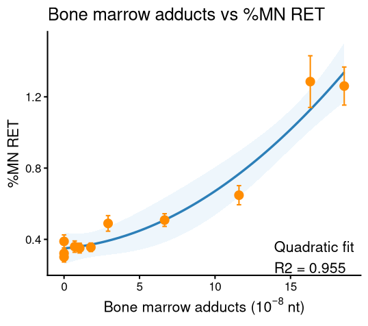
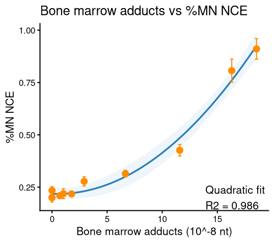
The highest quality study linking adducts to chromosomal aberrations (e.g. micronuclei) is an extensive study conducted by Long et al (2018). In this study, dose-concordance was extensively studied finding in bone marrow at a dose of 0.78 mg/kg bw/day there was a significant increase in DNA adducts without an increase in micronuclei formation until 3.13 mg/kg bw/day at which point there was a 4.5- and 3.7-fold increase in reticulocytes and monochromic erythrocytes, respectively. This study also shows that as the adduct levels increased, so do the micronuclei; when there is no increase in adduct formation at the lowest levels of exposure, there is no increase in micronuclei formation.
Mimmler et al. (2016) was able to establish both a dose and temporal concordance when they observed DNA adducts were detected as early as 2 hours after exposure to 50 uM PhIP and continued to increase through 48 hours. Following exposure to 1 uM N-OH-PhIP, adduct levels were detectable at 8 hours but declined by 24 hours. Chromosomal aberrations were assessed after a 24-hour exposure to 2.5 uM N-OH-PhIP followed by 14-hour mitotic arrest period, at which time aberration frequencies were significantly increased. The formation of aberrations is consistent with the initial increase of adduct formation over time after exposure establishing a time-response concordance as well as increasing dose resulting in increasing adduct formation and aberration formation.
Georgiadis et al. conducted multiple epidemiological studies on adduct and aberration formation in populations exposed to Environmental Tobacco Smoke (ETS) and B[a]P. In 2004 the researchers were able to link adduct formation, as a biomarker for exposure, to aberrant chromosomes, as a biomarker of effect. In 2005, they were able to find a ~2 to 2.5-fold increase in DNA adduct level with similar effects in aberrant chromosomes of individuals that are carriers of at least one polymorphism in the CYP1A1 gene compared to wild type allele. These findings support the essentiality of DNA adducts to chromosomal aberrations.
Table 1. Relevant empirical studies
| Species, Life Stage, Sex Tested | Stressor(s) | Specific Concordance | Effect on Upstream event (descriptive) | Effect on downstream event (descriptive) | Citation |
|---|---|---|---|---|---|
|
- MutaMouse - male - 12-13 weeks old |
B[a]P | Dose |
LOGEL for bone marrow: 0.78 mg/kg bw/day 50 mg/kg bw/day had a 19.6-fold increase from control |
Significant increase in RETs and NCEs at 3.13 mg/kg bw/day with RETs & NCEs having 4.5-fold and 3.7-fold increase respectively. | Long et al., 2018 |
|
- V79 chinese hamster cells (metabolically deficient) - V79 derived cells exposing human CP450 1A2 and human sulfotransferase 1A1, - Human colorectal cancer cells (HCT116, Caco-2) - Non-transformed human colonic epithelial cells (HCEC) |
PhIP & metabolite N-OH-PhIP | Dose & time |
Increase in adducts starting at 2 hours and increasing over the course of 48 hours from one exposure. At 1 uM dose, there was a peak of adducts after 8 hours with a decrease in adducts shown at 24 hours. When the dose was increased, (10-fold) the adduct level significantly increased without recovery at the 24-hour mark. |
After 24 hours, with a single dose of 2.5 uM N-OH-PhIP, there was a significantly slight increase in cells with aberrations. When the main repair system is inhibited and stressors are introduced, a significantly large increase in aberrations is observed. | Mimmler et al., 2016 |
| - Homo Sapiens (mean age 21.4 +- 1.5 yrs) | PAHs (Environmental Tobacco Smoke (ETS) & B[a]P) | Incidence & concentration | Higher ETS and PAH exposure, particularly in individuals with enhanced CYP1A1 genotypes, was associated with increased bulky DNA adduct levels | Increased frequencies of aberrant cells (chromosomal aberrations) were observed under the same exposure and genotype conditions that produced elevated bulky DNA adduct levels. Changes in chromosomal aberrations paralleled changes in DNA adduct levels |
Georgiadis et al., 2004 |
|
- Homo Sapiens - 18-25 yrs - 58 males - 136 females |
B[a]P | Incidence & concentration | Increase in bulky DNA adducts in lymphocytes | Increased amount of aberrant chromosomes | Georgiadis et al., 2005 |
Uncertainties and Inconsistencies
The empirical evidence supporting this KER primarily addresses dose concordance and incidence concordance with limited information regarding temporal concordance. This limitation reduces confidence in the quantitative characterization of the relationship. Additionally, without direct adduct measurement, it is difficult to determine downstream sensitivity or other experimental or biological factors. Overall, these limitations introduce uncertainty into the overall weight of evidence supporting the KER.
Known modulating factors
| Modulating Factor (MF) | MF Specification | Effect(s) on the KER | Reference(s) |
|---|---|---|---|
| Genetic Polymorphism | Any polymorphism impacting nucleotide excision repair, or double strand break repair | An increase in DNA adducts levels (KE upstream) and similar trends in aberrant chromosomes (KE downstream). | Georgiadis et al., 2004; Georgiadis et al., 2005 |
| Damaged Repair | Inhibition of ATR-interacting protein (recruits protein kinase ATR as key event in DNA damage response) | A significantly slight increase in aberrations (before inhibition) with a dramatic increase in ATR inhibition. | Mimmler et al., 2016 |
Quantitative Understanding of the Linkage
Available evidence suggests that a substantial increase in DNA adduct formation is required before detecting chromosomal aberrations. In the study conducted by Long et al., increases in DNA adducts were observed at doses as low as 0.20 mg/kg bw/day. While statistically significant increases in micronucleated reticulocytes and normochromatic erythrocytes were not observed until 3.13 mg/kg bw/day. Micronucleus frequencies increased approximately 4.5-fold and 3.7-fold above control levels at this dose, indicating that substantial accumulation of DNA adducts is required before measurable chromosomal damage is induced. This threshold-like relationship is further supported by benchmark dose modelling, where the BMD100 for DNA adduct formation (0.0286 mg/kg bw/day), is three orders of magnitude below the BMD for micronuclei (10.2 mg/kg bw/day), demonstrating that DNA adduct formation is a considerably more sensitive endpoint.
Evidence for the temporal relationship between these key events is more limited. Mimmler et al. demonstrated that DNA adducts were detectable within 2-8 hours of exposure and remained elevated for up to 48 hours depending on the compound and concentration used. In contrast, chromosomal aberrations were only evaluated after approximately 38 hours (24-hours exposure plus 14-hour mitotic arrest), at which point significant increases were observed. These findings indicate that DNA adduct formation precedes chromosomal aberration induction. However, the minimum duration or magnitude of adduct accumulation required to trigger chromosomal damage cannot be quantitatively defined because both key events were not assessed concurrently across multiple time points. Consequently, while there is strong evidence that DNA adduct formation occurs earlier and at lower doses than chromosomal aberrations, the precise temporal and quantitative relationship between the two key events remains uncertain. This shows that DNA adduct formation is a more sensitive endpoint and requires approximately 4- to 16-fold lower dose than those required to induce significant micronucleus formation.
Response-response Relationship
The graphs below were created using RStudio (version 4.6.1) and using AIC to find the models of best fit. Data was sourced from Long et al., 2018.

Time-scale
The ability to detail and develop a time-scale relationship for the two KEs is very complex and tissue-specific. Like in dose-response, each tissue has a different rate of metabolism, adduct formation, micronuclei formation, and repair. This causes a need for tissue specific timescales and not one larger organism wide timescale.
Known Feedforward/Feedback loops influencing this KER
No positive or negative feedback mechanisms related to this KER were identified in the literature reviewed. Consequently, there is currently no evidence to indicate whether feedback/feedforward loops influence this relationship, nor is there information available on their time-course, persistence, or homeostatic limits. It should be noted that this conclusion is based on the studies identified in the present literature search. Relevant publications may exist but were not captured due to limitations in search terminology, database coverage, or the number of papers assessed.
Domain of Applicability
Due to the conserved structure of DNA across cellular organisms, the potential for bulky adduct formation exists across taxa, sexes, and life stages. Bulky DNA adducts can form in a wide range of cell types following exposure to endogenous and exogenous genotoxic agents (Long et al., 2018). The cellular processes involved in DNA damage recognition and repair are also broadly conserved across species. While quantitative differences in adduct formation and repair may occur among taxa, sexes, and developmental stages, elevated levels of persistent DNA adduct increase the likelihood of chromosomal aberrations in dividing cells. Therefore, this KER is considered broadly applicable across taxa, sexes, and life stages.
References
Bi, Xiaohui, et al. “DNA Polymerase κ Is Specifically Required for Recovery from the Benzo[a]Pyrene-Dihydrodiol Epoxide (BPDE)-Induced S-Phase Checkpoint.” The Journal of Biological Chemistry [United States], vol. 280, no. 23, June 2005, pp. 22343–55, https://doi.org/10.1074/jbc.M501562200.
Georgiadis, P., et al. “Impact of Phase I or Phase II Enzyme Polymorphisms on Lymphocyte DNA Adducts in Subjects Exposed to Urban Air Pollution and Environmental Tobacco Smoke.” Toxicology Letters [Shannon], vol. 149, no. 1, April 2004, pp. 269–80, https://doi.org/10.1016/j.toxlet.2003.12.038.
Georgiadis, Panagiotis, et al. “Interactions between CYP1A1 Polymorphisms and Exposure to Environmental Tobacco Smoke in the Modulation of Lymphocyte Bulky DNA Adducts and Chromosomal Aberrations.” Carcinogenesis (New York) [Oxford], vol. 26, no. 1, January 2005, pp. 93–101, https://doi.org/10.1093/carcin/bgh294.
Kaufmann, William K. “Pathways of Human Cell Post-Replication Repair.” Carcinogenesis (New York) [Oxford], vol. 10, no. 1, January 1989, pp. 1–11, https://doi.org/10.1093/carcin/10.1.1.
Long, Alexandra S., et al. “Benchmark Dose Analyses of Multiple Genetic Toxicity Endpoints Permit Robust, Cross-Tissue Comparisons of MutaMouse Responses to Orally Delivered Benzo[a]Pyrene.” Archives of Toxicology [Berlin/Heidelberg], vol. 92, no. 2, February 2018, pp. 967–82, https://doi.org/10.1007/s00204-017-2099-2.
Mimmler, Maximilian, et al. “DNA Damage Response Curtails Detrimental Replication Stress and Chromosomal Instability Induced by the Dietary Carcinogen PhIP.” Nucleic Acids Research [England], vol. 44, no. 21, December 2016, pp. 10259–76, https://doi.org/10.1093/nar/gkw791.
Nikolova, Teodora, et al. “The γH2AX Assay for Genotoxic and Nongenotoxic Agents: Comparison of H2AX Phosphorylation with Cell Death Response.” Toxicological Sciences [United States], vol. 140, no. 1, July 2014, pp. 103–17, https://doi.org/10.1093/toxsci/kfu066.
Pavel Rossner, et al. “Genetic Toxicology and Carcinogenesis.” Toxicology for the Health and Pharmaceutical Sciences, edited by Antonio Peña-Fernández et al., 1st ed., CRC Press, 2022, pp. 81–97, https://doi.org/10.1201/9780203730584-5.
Rossner, Pavel, et al. “Molecular Epidemiology Focused on Airborne Carcinogens.” Air Pollution and Health Effects, edited by Srikanth S. Nadadur and John W. Hollingsworth, Springer London, Limited, 2015, pp. 185–212, https://doi.org/10.1007/978-1-4471-6669-6_7.
Tu, Lan Chun, et al. “Hedamycin, a DNA Alkylator, Induces γH2AX and Chromosome Aberrations: Involvement of Phosphatidylinositol 3-Kinase–Related Kinases and DNA Replication Fork Movement.” Molecular Cancer Therapeutics, vol. 4, no. 8, August 2005, pp. 1175–85, https://doi.org/10.1158/1535-7163.MCT-05-0054.