This AOP is licensed under the BY-SA license. This license allows reusers to distribute, remix, adapt, and build upon the material in any medium or format, so long as attribution is given to the creator. The license allows for commercial use. If you remix, adapt, or build upon the material, you must license the modified material under identical terms.
AOP: 505
Title
Reactive Oxygen Species (ROS) formation leads to cancer via inflammation pathway
Short name
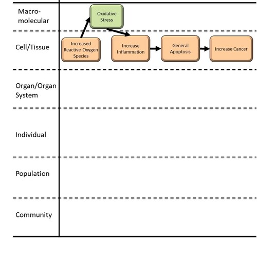
Graphical Representation

Point of Contact
Contributors
- John Frisch
Coaches
OECD Information Table
| OECD Project # | OECD Status | Reviewer's Reports | Journal-format Article | OECD iLibrary Published Version |
|---|---|---|---|---|
This AOP was last modified on October 30, 2024 15:12
Revision dates for related pages
| Page | Revision Date/Time |
|---|---|
| Increase, Reactive oxygen species | June 12, 2025 01:27 |
| Increase, Oxidative Stress | February 11, 2026 07:05 |
| Increase, Inflammation | February 28, 2024 06:33 |
| General Apoptosis | October 18, 2023 12:20 |
| Increase, Cancer | August 22, 2023 14:32 |
| Increase, ROS leads to Increase, Oxidative Stress | August 02, 2024 15:40 |
| Increase, Oxidative Stress leads to Increase, Inflammation | October 19, 2023 09:39 |
| Increase, Inflammation leads to General Apoptosis | October 19, 2023 09:41 |
| General Apoptosis leads to Increase, Cancer | October 19, 2023 09:46 |
| Polyethylene AS low Mol.Wt. | July 31, 2023 09:43 |
| Polyvinyl chloride | July 31, 2023 09:45 |
Abstract
Reactive oxygen species (ROS) are derived from oxygen molecules and can occur as free radicals (ex. superoxide, hydroxyl, peroxyl) or non-radicals (ex. ozone, singlet oxygen). ROS production occurs via a variety of normal cellular process; however, in stress situations (ex. exposure to radiation, chemical or biological stressors) reactive oxygen species levels dramatically increase and cause damage to cellular components. In this Adverse Outcome Pathway (AOP) we focus on the inflammation response to increases in oxidative stress. Inflammation pathways include a molecular response (ex. interleukins, cytokines, interferons) and produces visible tissue swelling during histology examinations. In this AOP we focus on the apoptosis response to cellular damage. Pathways leading to apoptosis, or single cell death, have traditionally been studied as both independent and simultaneous from pathways leading to necrosis, or tissue-wide cell death, with both overlap and distinct mechanisms (Elmore 2007). For the purposes of this AOP, we are characterizing cancer due to widespread cell-death, and recognize the complications in separating the related apoptosis and necrosis pathways.
AOP Development Strategy
Context
This Adverse Outcome Pathway (AOP) focuses on the pathway in which an established molecular disruption, increased levels of reactive oxygen species (ROS), leads to increased cancer through inflammation and cell/death/apoptosis. Environmental stressors leading to increased reactive oxygen species result in a variety of stress responses, visible through inflammation. These stress responses have been studied in many eukaryotes, including mammals (humans, lab mice, lab rats), teleost fish, and invertebrates (cladocerans, mussels).
Strategy
This AOP was developed as part of an Environmental Protection Agency effort to represent putative AOPs from peer-reviewed literature which were heretofore unrepresented in the AOP-Wiki. Jeong and Choi (2020) and Jeong and Choi (2019) provided initial network analysis from microplastic stressors, guided by weight of evidence from ToxCast assays. These publication, and the work cited within, were used create and support this AOP and its respective KE and KER pages.
The AOP-wiki authors did a further evaluation of published peer-reviewed literature to provide additional evidence in support of the AOP. A companion adverse outcome pathway is planned for an additional pathway initiated by reactive oxygen species (ROS), leading to increased cancer: Decreased, PPARalpha transactivation of gene expression leads to Alteration, lipid metabolism.
Summary of the AOP
Events:
Molecular Initiating Events (MIE)
Key Events (KE)
Adverse Outcomes (AO)
| Type | Event ID | Title | Short name |
|---|
| MIE | 1115 | Increase, Reactive oxygen species | Increase, ROS |
| KE | 1392 | Increase, Oxidative Stress | Increase, Oxidative Stress |
| KE | 149 | Increase, Inflammation | Increase, Inflammation |
| KE | 1513 | General Apoptosis | General Apoptosis |
| AO | 885 | Increase, Cancer | Increase, Cancer |
Relationships Between Two Key Events (Including MIEs and AOs)
| Title | Adjacency | Evidence | Quantitative Understanding |
|---|
| Increase, ROS leads to Increase, Oxidative Stress | adjacent | High | Not Specified |
| Increase, Oxidative Stress leads to Increase, Inflammation | adjacent | High | Not Specified |
| Increase, Inflammation leads to General Apoptosis | adjacent | High | Not Specified |
| General Apoptosis leads to Increase, Cancer | adjacent | High | Not Specified |
Network View
Prototypical Stressors
Life Stage Applicability
| Life stage | Evidence |
|---|---|
| All life stages | High |
Taxonomic Applicability
Sex Applicability
| Sex | Evidence |
|---|---|
| Unspecific | High |
Overall Assessment of the AOP
|
1. Support for Biological Plausibility of Key Event Relationships: Is there a mechanistic relationship between KEup and KEdown consistent with established biological knowledge? |
|
|
Key Event Relationship (KER) |
Level of Support Strong = Extensive understanding of the KER based on extensive previous documentation and broad acceptance. |
|
Relationship 2009: Increased, Reactive oxygen species leads to Oxidative Stress |
Strong support. The relationship between increases in reactive oxygen species and oxidative stress is broadly accepted and consistently supported across taxa. |
|
Relationship 2975: Oxidative Stress leads to Increase, Inflammation |
Strong support. The relationship between oxidative stress and increased inflammation is established. |
|
Relationship 2976: Increase, Inflammation leads to General Apoptosis |
Strong support. The relationship between increased inflammation and general apoptosis is established. Inflammation has been shown as an initiating event for activation of apoptosis; arguably more studies have been conducted linking inflammation to necrosis pathways. |
|
Relationship 2977: General Apoptosis leads to Increase, Cancer |
Strong support. The relationship between failure of apoptosis pathways to initiate cell death pathways and increases in cancer is broadly accepted and consistently supported across taxa. |
|
Overall |
Strong support. Extensive understanding of the relationships between events from empirical studies from a variety of taxa. |
Domain of Applicability
Life Stage: The life stage applicable to this AOP is all life stages. Older individuals are more likely to manifest this adverse outcome pathway (adults > juveniles > embryos) due to accumulation of reactive oxygen species.
Sex: This AOP applies to both males and females.
Taxonomic: This AOP appears to be present broadly, with representative studies including mammals (humans, lab mice, lab rats), teleost fish, and invertebrates (cladocerans, mussels).
Essentiality of the Key Events
Support for the essentiality of the key events can be obtained from a wide diversity of taxonomic groups, with mammals (lab ice, lab rats, human cell lines), telost fish, and invertebrates (cladocerans and mussels) particularly well-studied.
|
2. Essentiality of Key Events: Are downstream KEs and/or the AO prevented if an upstream KE is blocked? |
|
|
Key Event (KE) |
Level of Support Strong = Direct evidence from specifically designed experimental studies illustrating essentiality and direct relationship between key events. Moderate = Indirect evidence from experimental studies inferring essentiality of relationship between key events due to difficulty in directly measuring at least one of key events. |
|
MIE 1115: Increased, Reactive oxygen species |
Strong support. Increased Reactive oxygen species (ROS) levels are a primary cause of oxidative stress. Evidence is available from studies of stressor exposure and resulting changes in gene expression and protein/enzyme levels. |
|
KE 1392: Oxidative Stress |
Strong support. Oxidative stress is a cause of inflammation. Evidence is available from studies of stressor exposure and resulting changes in gene expression, protein/enzyme levels, and histology. |
|
KE 149: Increase, Inflammation |
Strong support. Inflammation is a cause of apoptosis. Evidence is available from studies of stressor exposure and resulting changes in gene expression, protein/enzyme levels, and histology. |
|
KE 1513: General Apoptosis |
Moderate support. Failure of apoptosis allows cancer cells to proliferate. Evidence is available from studies of stressor exposure and resulting changes in gene expression, protein/enzyme levels, and histology. |
|
AO 885: Increase, Cancer |
Strong support. Cancer proliferates due to a variety of stressors and breakdown of multiple cellular processes. Evidence is available from studies of stressor exposure and resulting changes in gene expression, protein/enzyme levels, and histology. |
|
Overall |
Moderate to strong support. Direct evidence from empirical studies for most key events, with more inferential evidence rather than direct evidence for apoptosis. |
Evidence Assessment
|
Path |
Support |
|
Increased, Reactive oxygen species leads to Oxidative Stress |
Biological plausibility is high. Representative studies have been done with mammals (Liu et al. 2015; Deng et al. 2017; Schrinzi et al. 2017; Jeong and Choi 2020); fish (Oliveira et al. 2013; Lu et al. 2016; Alomar et al. 2017; Chen et al. 2017; Veneman et al. 2017; Barboza et al. 2018; Choi et al. 2018; Espinosa et al. 2018); invertebrates (Browne et al. 2013; Avio et al. 2015; Jeong et al. 2016, 2017; Paul-Pont et al. 2016; Imhof et al. 2017; Lei et al. 2018; Yu et al. 2018). |
|
Oxidative Stress leads to Increase, Inflammation |
Biological plausibility is high. Representative studies have been done with mammals (Gamo et al. 2008; Jeong and Choi 2020); fish (Lu et al. 2016; Jin et al. 2018); invertebrates (Lei et al. 2018). For review (Wright and Kelly 2017). |
|
Increase, Inflammation leads to General Apoptosis |
Biological plausibility is high. Representative studies have been done with mammals (Gamo et al. 2008); fish (Karami et al. 2016; Lu et al. 2016; Jin et al. 2018). For review (Balkwill 2003, Villeneuve et al. 2018). |
|
General Apoptosis leads to Increase, Cancer |
Biological plausibility is high. Representative studies have been done with mammals (Pavet et al. 2014; Jeong and Choi 2020). For review (Heinlein and Chang 2004; Vihervaara and Sistonen 2014). |
|
3. Empirical Support for Key Event Relationship: Does empirical evidence support that a change in KEup leads to an appropriate change in KEdown? |
|
|
Key Event Relationship (KER) |
Level of Support Strong = Experimental evidence from exposure to toxicant shows consistent change in both events across taxa and study conditions. |
|
Relationship 2009: Increased, Reactive oxygen species leads to Oxidative Stress |
Strong support. Increases in ROS lead to increases in oxidative stress, primarily from studies examining responses in enzyme and gene levels for enzymes that catalyze reactions that reduce ROS levels. |
|
Relationship 2975: Oxidative Stress leads to Increase, Inflammation |
Strong support. Increases in oxidative stress leads to increases in inflammation, primarily from histology studies measuring tissue swelling, and increases in gene levels for proinflammatory mediators. |
|
Relationship 2976: Increase, Inflammation leads to General Apoptosis |
Strong support. Increases in inflammation leads to apoptosis, primarily from studies of increased gene expression of tumor necrosis factor. |
|
Relationship 2977: General Apoptosis leads to Increase, Cancer |
Strong support. Mechanistic studies show that failure for apoptosis to eliminate cancer cells allows increases in cancer proliferation. |
|
Overall |
Strong support. Evidence from empirical studies shows consistent change in both events from a variety of taxa |
For overview of the biological mechanisms involved in this AOP, see Liu et al. (2015) and Jeong and Choi (2020); their studies analyzed ToxCast in vitro assays of mammalian acute toxicity data to identify correlations between toxicity pathways and chemical stressors, providing support for the key event relationships represented here.
Known Modulating Factors
| Modulating Factor (MF) | Influence or Outcome | KER(s) involved |
|---|---|---|
Quantitative Understanding
Considerations for Potential Applications of the AOP (optional)
References
Alomar, C., Sureda, A., Capo, X., Guijarro, B., Tejada, S. and Deudero, S. 2017. Microplastic ingestion by Mullus surmuletus Linnaeus, 1758 fish and its potential for causing oxidative stress. Environmental Research 159: 135-142.
Avio, C.G., Gorbi, S., Milan, M., Benedetti, M., Fattorini, D., D’Errico, G., Pauletto, M., Bargelloni, L., and Regoli, F. 2015. Pollutants bioavailability and toxicological risk from microplastics to marine mussels. Environmental Pollutants 198: 211-222.
Barboza, LG.A., Vieira, L.R., Branco, V., Figueiredo, N., Carvalho, F., Carvalho, C., and Guilhermino, L. 2018. Microplastics cause neurotoxicity, oxidative damage and energy-related changes and interact with the bioaccumulation of mercury in the European seabass, Dicentrachus labrux (Linneaeus, 1758). Aquatic Toxicology 195: 49-57.
Balkwill, F. 2003. Chemokine biology in cancer. Seminars in Immunology 15: 49-55.
Browne, M.A. Niven, S.J., Galloway, T.S., Rowland, S.J., and Thompson, R.C. 2013. Microplastic moves pollutants and additives to worms, reducing functions linked to health and biodiversity. Current Biology 23: 2388-2392.
Chen, Q., Gundlach, M., Yang, S., Jiang, J., Velki, M., Yin, D., and Hollert, H. 2017 Quantitative investigation of the mechanisms of microplastics and nanoplastics toward larvae locomotor activity. Science of the Total Environment 584-585: 1022-1031.
Choi, J.S., Jung, Y.J., Hong, N.H., Hong, S.H., and Park, J.W. 2018. Toxicological effects of irregularly shaped and spherical microplastics in a marine teleost, the sheepshead minnow (Cyprinodon variegatus). Marine Pollution Bulletin 129: 231-240.
Deng, Y., Zhang, Y., Lemos, B., and Ren, H. 2017. Tissue accumulation of microplastics in mice and biomarker responses suggest widespread health risks of exposure. Science Reports 7: 1-10.
Elmore, S. 2007. Apoptosis: A Review of Programmed Cell Death. Toxicologic pathology 35 (4): 495-516.
Espinosa, C., Garcia Beltran, J.M., Esteban, M.A., and Cuesta, A. 2018. In vitro effects of virgin microplastics on fish head-kidney leucocyte activities. Environmental Pollution 235: 30-38.
Gamo, K., Kiryu-Seo, S., Konishi, H., Aoki, S., Matushima, K., Wada, K., and Kiyama, H. 2008. G-protein-coupled receptor screen reveals a role for chemokine recepteor CCR5 in suppressing microglial neurotoxicity. Journal of Neuroscience 28: 11980-11988.
Heinlein, C.A. and Chang, C. 2004. Androgen receptor in prostate cancer. Endocrine Reviews 25: 276-308.
Imhof, H.K., Rusek, J., Thiel, M., Wolinska, J., and Laforsch, C. 2017. Do microplastic particles affect Daphnia magna at the morphological life history and molecular level? Public Library of Science One 12: 1-20.
Jeong, J. and Choi, J. 2019. Adverse outcome pathways potentially related to hazard identification of microplastics based on toxicity mechanisms. Chemosphere 231: 249-255.
Jeong, J. and Choi, J. 2020. Development of AOP relevant to microplastics based on toxicity mechanisms of chemical additives using ToxCast™ and deep learning models combined approach. Environment International 137:105557.
Jeong, C.B., Kang, H.M., Lee, M.C., Kim, D.H., Han, J., Hwang, D.S. Souissi, S., Lee, S.J., Shin, K.H., Park, H.G., and Lee, J.S. 2017. Adverse effects of microplastics and oxidative stress-induced MAPK/NRF2 pathway-mediated defense mechanisms in the marine copepod Paracyclopina nana. Science Reports 7: 1-11.
Jeong, C.B., Wong, E.J., Kang, H.M., Lee, M.C., Hwang, D.S., Hwang, U.K., Zhou, B., Souissi, S., Lee, S.J., and Lee, J.S. 2016. Microplastic size-dependent toxicity, oxidative stress induction, and p-JNK and p-p38 activation in the Monogonout rotifer (Brachionus koreanus). Environmental Science and Technology 50: 8849-8857.
Jin, Y., Xia, J., Pan, Z., Yang, J., Wang, W., and Fu, Z. 2018. Polystyrene microplastics induce microbiota dysbiosis and inflammation in the gut of adult zebrafish. Environmental Pollution 235: 322-329.
Karami, A., Romano, N., Galloway, T. and Hamzah, H. 2016. Virgin microplastics cause toxicity and modulate the impacts of phenanthrene on biomarker responses in African catfish (Clarias gariepinus). Environmental Research 151: 58-70.
Lei, L., Wu, S., Lu, S., Liu, M., Song, Y., Fu, Z., Shi, H., Raley-Susman, K.M., and He, D. 2018. Microplastic particles cause intestinal damage and other adverse effects in zebrafish Danio rerio and nematode Caenorhabditis elegans. Science of the Total Environment 619-620: 1-8.
Liu, J., Mansouri, K., Judson, R.S., Martin, M.T., Hong, H., Chen, M., Xu, X., Thomas, R.S., and Shah, I. 2015. Predicting hepatoxicity using ToxCast in vitro bioactivity and chemical structure. Chemical Research in Toxicology 28: 738-751.
Lu, Y., Zhang, Y., Dengy, Y., Jiang, W., Zhao, Y., Geng, J., Ding, L., Ren, H. 2016. Uptake and accumulation of polystyrene microplastics in zebrafish (Danio rerio) and toxic effects in liver. Environmental Science and Technology 50: 4054-4060.
Oliveira, M., Ribeiro, A., Hylland, K., and Guilhermino, L. 2013. Single and combined effects of microplastics and pyrene on juveniles (0+ group) of the common goby Pomatoschistus microps (Teleostei, Gobiidae). Ecological Indicators 34: 641-647.
Paul-Pont, I., Lacroix, C., Gonzalez Fernandez, D., Hegaret, H., Lambert, C., Le Goic, N., Frere, L., Cassone, A.L., Sussarellu, R. Fabioux, C., Guyomarch, J., Albentosa, M., Huvet, A., and Soudant, P. 2016. Exposure of marine mussels Mytillus spp. to polystyrene microplastics: Toxicity and influence on fluoranthene bioaccumulation. Environmental Pollution 216: 724-737.
Pavet, V., Shlyakhtina, Y., He, T., Ceschin, D.G., Kohonen, P., Perala, M., Kallioniemi, O., and Gronemeyer, H. 2014. Plasminogen activator urokinase expression reveals TRAIL responsiveness and support fractional survival of cancer cells. Cell Death and Disease 5: e1043.
Schrinzi, G.F., Perez-Pomeda, I., Sanchis, J., Rossini, C., Farre, M., and Barcelo, D. 2017. Cytotoxic effects of commonly used nanomaterials and microplastics on cerebral and epithelial human cells. Environmental Research 159: 579-587.
Veneman, W.J., Spaink, H.P., Brun, N.R., Bosker, T., and Vijver, M.G. 2017. Pathway analysis of systemic transcriptome responses to injected polystyrene particles in zebrafish larvae. Aquatic Toxicology 190: 112-120.
Vihervaara, A. and Sistonen, L. 2014. HSF1 at a glance. Journal of Cell Scientce 127: 261-266.
Villeneuve, D.L., Landesmann, B., Allavena, P., Ashley, N., Bal-Price, A., Corsini, E., Halappanavar, S., Hussell, T., Laskin, D., Lawrence, T., Nikolic-Paterson, D., Pallary, M., Paini, A., Pietrs, R., Roth, R., and Tschudi-Monnet, F. 2018. Toxicological Sciences 346:352.
Wright, S.L. and Kelly, F.J. 2017. Plastic and human health: a micro issue? Enviromental Science and Technology 51: 6634-6647.
Yu, P., Liu, Z., Wu, D., Chen, M., Lv, W., and Zhao, Y. 2018. Accumulation of polystyrene microplastics in juvenile Eriocheir sinensis and oxidative stress effects in the liver. Aquatic Toxicology 200: 28-36.