
The authors have designated this AOP as all rights reserved. Re-use in any form requires advanced permission from the authors.
AOP: 547
Title
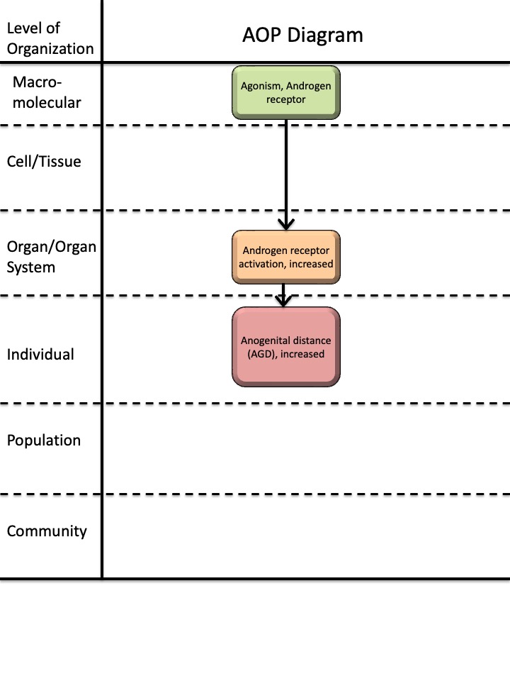
Androgen receptor agonism leading to long anogenital distance (AGD) in female offspring
Short name
Graphical Representation
Additional AOP Exploration Options
Click links below to explore AOP 547, Androgen receptor agonism leading to long anogenital distance (AGD) in female offspring in tools offered by third parties.

Point of Contact
Contributors
- Johanna Zilliacus
Coaches
OECD Information Table
| OECD Project # | OECD Status | Reviewer's Reports | Journal-format Article | OECD iLibrary Published Version |
|---|---|---|---|---|
This AOP was last modified on August 30, 2026 05:59
Revision dates for related pages
| Page | Revision Date/Time |
|---|---|
| Androgen receptor activation, increased | November 02, 2024 09:53 |
| Agonism, Androgen receptor | November 05, 2024 07:49 |
| Anogenital distance (AGD), increased | August 30, 2026 06:23 |
| Agonism, Androgen receptor leads to Androgen receptor activation, increased | November 24, 2024 07:47 |
| Androgen receptor activation, increased leads to Anogenital distance (AGD), increased | August 30, 2026 06:00 |
| 17-Methyltestosterone | August 30, 2026 05:46 |
| Testosterone propionate | August 30, 2026 05:48 |
| 17beta-Trenbolone | November 29, 2016 18:42 |
Abstract
The purpose of developing this AOP was to establish a framework for identifying endocrine disruptors based on the measurement of anogenital distance (AGD) in female rodents. AGD is defined as the distance between the anus and the external genitalia. In both rodents and humans, female AGD is approximately half the length of male AGD (Liu et al., 2014; Salazar-Martinez et al., 2004; Schwartz et al., 2019; Sharpe, 2020; Thankamony et al., 2016; Wise, 2024). This difference reflects androgen-dependent regulation of perineal growth during fetal development in males, whereas in females, the absence of androgens limits this growth (Ipulan et al., 2016; Schwartz et al., 2019; Sharpe, 2020; Welsh et al., 2008).
AGD is considered an apical endpoint and is included in OECD test guidelines TG414, TG416, TG421, TG422 and TG443 (OECD, 2001; OECD, 2018; OECD, 2025a; OECD, 2025b; OECD, 2025c). According to ECHA/EFSA guidance for identifying endocrine disruptors, AGD is classified as an EATS-mediated parameter and is used in the identification of endocrine disruptors (ECHA/EFSA, 2018).
The molecular initiating event (MIE) for this AOP is androgen receptor (AR) agonism, meaning the binding of a compound to the AR that activates the receptor. This leads to the first key event (KE): increased AR activation in tissues and organs in vivo, including the perineum. During fetal development in females, increased AR activation in the perineum results in increased AGD considered an adverse outcome (AO).
The overall weight of evidence supporting this AOP is high. Biological plausibility for both key event relationships (KERs) is strong, as androgen receptor mechanisms and androgenic effects on AGD are well established. Empirical evidence for the KERs is based on studies showing that AR agonists increase female AGD in prenatal rat studies.
This AOP is applicable for identifying androgenic endocrine disruptors.
AOP Development Strategy
Context
This AOP was developed as part of the European Food Safety Authority (EFSA) project (Grant Agreement No. GP/EFSA/PREV/2022/01) aimed at creating AOPs for endocrine disruptors. The purpose of this AOP is to provide a framework for identifying endocrine disruptors associated with increased AGD in female rodent offspring. Other AOPs within the project address shortened AGD in males (AOPs 305, 306, and 307).
The molecular initiating event (MIE25: Agonism, Androgen receptor) and key event (KE2274: Androgen receptor activation, increased) described in this AOP can serve as a foundation for developing additional AOPs related to adverse outcomes resulting from androgen receptor agonism.
Furthermore, this AOP can be expanded into an AOP network by incorporating pathways such as “Increased testosterone levels leading to long anogenital distance in female offspring” and “Increased dihydrotestosterone levels leading to long anogenital distance in female offspring.” These can be developed using the key events (KE2272, KE2273) and key event relationships (KER3380, KER3381) established in the EFSA-funded project.
A shortened AGD is recognized as a sensitive marker of reduced AR activity during the masculinization programming window in the male, whereas in the female a longer AGD is a marker of increased AR activity and masculinization of the female fetus in this period.
Strategy
This AOP was developed in two steps.
First, an upstream AOP network for “increased androgen activity” was constructed. The AOP-Wiki was screened to identify existing key events (KEs) related to increased androgen activity by searching KE and KER pages for the terms androgen, testosterone, dihydrotestosterone, and DHT. Two relevant KEs were identified, but no relevant KERs. KE25 was updated, while KE286 required no revision. All other KEs and KERs were newly developed following the methodology outlined in the AOP Developers’ Handbook (version 2.7): KE2272, KE2273, KE2274, KER3378, KER3379, KER3380, and KER3381.
The network reflects processes widely regarded as canonical, and evidence was primarily drawn from review articles and book chapters identified through searches in PubMed and library catalogues. For the current AOP, the following KEs from the upstream network were used: KE25, KE2274, and KER3378. The remaining KEs and KERs could be used to expand the current AOP into an AOP network as described above.
In the second step, KE2365 and KER3628 were developed.
Evidence for KE2365 was drawn mainly from review articles identified through PubMed searches. A systematic weight-of-evidence approach was applied to collect, evaluate, extract, and integrate evidence for KER3628, as detailed on its dedicated page. Briefly, a scoping literature search was first conducted in PubMed to identify relevant model substances. Based on these findings, a targeted search was performed in PubMed and Web of Science using terms related to the model substances and AGD.
Study reliability for included publications was assessed using the Science in Risk Assessment and Policy (SciRAP) tools for in vivo studies (http://www.scirap.org), and overall confidence in the evidence for each model substance was categorized.
The AOP includes three KEs: the MIE, the AO, and one intermediate KE.
These were selected to capture the main steps in the pathway from AR agonism to increased AGD. Additional KEs could be added, such as KE286 (Altered transcription of genes by the AR) and a KE describing androgen regulation of perineal growth, which likely involves androgen-mediated development of the levator ani and bulbocavernosus (LABC) muscle complex in the perineum. However, sufficient empirical evidence for these related KERs is currently lacking.
Summary of the AOP
Events:
Molecular Initiating Events (MIE)
Key Events (KE)
Adverse Outcomes (AO)
| Type | Event ID | Title | Short name |
|---|
| MIE | 25 | Agonism, Androgen receptor | Agonism, Androgen receptor |
| KE | 2274 | Androgen receptor activation, increased | Androgen receptor activation, increased |
| AO | 2365 | Anogenital distance (AGD), increased | Anogenital distance (AGD), increased |
Relationships Between Two Key Events (Including MIEs and AOs)
| Title | Adjacency | Evidence | Quantitative Understanding |
|---|
| Agonism, Androgen receptor leads to Androgen receptor activation, increased | adjacent | High | Low |
| Androgen receptor activation, increased leads to Anogenital distance (AGD), increased | adjacent | High | Low |
Network View
Prototypical Stressors
Life Stage Applicability
| Life stage | Evidence |
|---|---|
| Fetal | High |
Taxonomic Applicability
Sex Applicability
| Sex | Evidence |
|---|---|
| Female | High |
Overall Assessment of the AOP
Domain of Applicability
Life stage applicability
The life stage applicability domain for this AOP is fetal. Although AR activation occurs during fetal development, puberty, and adulthood, the regulation of AGD growth is specifically controlled by androgens during the masculinization programming window, which takes place in fetal development (Dalton & Gao, 2010; Luetjens & Weinbauer, 2012; Naamneh Elzenaty et al., 2022; Sutinen et al., 2017; Welsh et al., 2008).
Taxonomic applicability
The biologically plausible taxonomic applicability domain for this AOP is mammals, as fetal masculinization is regulated by androgens across all mammalian species (Welsh et al., 2014). The empirical taxonomic applicability domain is rat and human, supported by prenatal studies in rat demonstrating that androgen receptor activation leads to a measurable increase in female offspring AGD (Armoskus et al., 2014; Guerra et al., 2014; Hotchkiss et al., 2007; Sathishkumar et al., 2011; Welsh et al., 2009; Wilson et al., 2002; Wolf et al., 2002; Wolf et al., 2004) and epidemiological studies suggesting that elevated androgen levels in women are associated with increased AGD (Mira-Escolano et al., 2014; Pan et al., 2021; Zamani et al., 2023).
Sex applicability
The sex applicability domain for this AOP is females, supported by studies showing effects of androgen activation on female AGD (Armoskus et al., 2014; Guerra et al., 2014; Hotchkiss et al., 2007; Mira-Escolano et al., 2014; Pan et al., 2021; Sathishkumar et al., 2011; Welsh et al., 2009; Wilson et al., 2002; Wolf et al., 2002; Wolf et al., 2004; Zamani et al., 2023).
Essentiality of the Key Events
|
KE |
Evidence |
Level of evidence |
|
MIE25 |
Direct evidence on effect on KE2274: Studies in AR knockout mice demonstrate that AR is essential for mediating effects on the reproductive system by AR activation (De Gendt et al., 2004; Holdcraft & Braun, 2004; Matsumoto et al., 2003; Notini et al., 2005; Yeh et al., 2002). AR antagonists flutamide, procymidone and vinclozolin inhibit AR activity in vivo as shown in the OECD test guideline Hershberger assay (Browne et al., 2018). Direct evidence on effect on AO2365: Studies in AR knockout mice demonstrate that AR is essential for a longer AGD, as male AR knockout mice exhibit AGD lengths comparable to female wild-type mice (Yeh et al., 2002, Sato et al., 2004). It should be noted AGD in male mice was studied but the AO2365 is in females. Prenatal exposure to the AR antagonists flutamide, procymidone and vinclozolin results in shorter AGD in rat studies (Casto et al., 2003; Foster & Harris, 2005; Fussell et al., 2015; Goto et al., 2004; Gray et al., 1994; Hass et al., 2007; Hass et al., 2012; Inawaka et al., 2010; Kita et al., 2016; Ostby et al., 1999; McIntyre et al., 2001; Shimamura et al., 2002; Wolf et al., 2000; Yamasaki et al., 2005). It should be noted AGD in male rats was studied but the AO2365 is in females. |
High. There is direct evidence from knockout and antagonist studies. There are no inconsistencies identified but it should be noted that the effect on AGD was studied in male animals. |
|
KE2274 |
Direct evidence on effect on AO2365: Studies in AR knockout mice demonstrate that AR activity is essential for a longer AGD, as male AR knockout mice exhibit AGD lengths comparable to female wild-type mice (Yeh et al., 2002, Sato et al., 2004). It should be noted AGD in male mice was studied but the AO2365 is in females. Prenatal exposure to the AR antagonists flutamide, procymidone and vinclozolin results in reduced AGD in rat studies (Casto et al., 2003; Foster & Harris, 2005; Fussell et al., 2015; Goto et al., 2004; Gray et al., 1994; Hass et al., 2007; Hass et al., 2012; Inawaka et al., 2010; Kita et al., 2016; Ostby et al., 1999; McIntyre et al., 2001; Shimamura et al., 2002; Wolf et al., 2000; Yamasaki et al., 2005). It should be noted AGD in male rats was studied but the AO2365 is in females. |
High. There is direct evidence from knockout and antagonist studies. There are no inconsistencies identified, but it should be noted that the effect on AGD was studied in male animals. |
Evidence Assessment
Assessment of biological plausibility
|
KER |
Assessment |
|
KER 3378: Agonism, Androgen receptor leads to Androgen receptor activation, increased |
The biological plausibility of the KER is High. It is a generally recognised process, canonical knowledge, that binding of agonists to AR leads to increased AR activation in vivo (Dalton & Gao, 2010; Luetjens & Weinbauer, 2012; Naamneh Elzenaty et al., 2022; Sutinen et al., 2017). |
|
KER 3628: Androgen receptor activation, increased leads to AGD, increased |
The biological plausibility of the KER is High. It is a well-documented by numerous studies in rats that AR activation drives the fetal masculinization process, including the growth of the perineum, which can be measured as AGD (Liu et al., 2014; Salazar-Martinez et al, 2004; Schwartz et al., 2019; Sharpe, 2020; Thankamony et al., 2016; Welsh et al., 2008; Wise, 2024). This is supported by epidemiological studies (Mira-Escolano et al., 2014; Pan et al., 2021; Zamani et al., 2023). |
Assessment of empirical evidence
|
KER |
Assessment |
|
KER 3378: Agonism, Androgen receptor leads to Androgen receptor activation, increased |
The empirical support for the KER is High. AR agonists such as testosterone propionate, methyl testosterone and trenbolone have been identified as agonists in the US EPA Androgen receptor pathway model, which integrates in vitro assays for receptor binding, coregulator recruitment and transactivation. These stressors have also been shown to increase androgen activity in Hershberger assay and other in vivo assays (Kleinstreuer et al., 2018; Browne et al., 2018). Assessing dose and temporal concordance for this KER is challenging because the upstream KE is measured in vitro, whereas the downstream KE reflects on increased in vivo activation. There are no available data on incidence concordance. |
|
KER 3628: Androgen receptor activation, increased leads to AGD, increased |
The empirical support for the KER is High. The empirical evidence is based on four model substances: methyl testosterone, testosterone, testosterone propionate and trenbolone. These substances bind AR and act as AR agonists, as demonstrated in in vitro studies (Kleinstreuer et al., 2018; Judson et al., 2020) and androgen activity in in vivo Hershberger assays (Browne et al., 2018). Prenatal exposure to these substances in rat studies has been shown to increase AGD in female offspring, as reported in multiple studies (Armoskus et al., 2014; Guerra et al., 2014; Hotchkiss et al., 2007; Juarez et al., 1995; Kawashima et al., 1975; McCoy et al., 1992; Rhees et al., 1997; Tehrani et al., 2014; Welsh et al., 2009; Wilson et al., 2002; Wolf et al., 2002; Wolf et al., 2004; Wu et al., 2010) Assessing dose and temporal concordance for this KER is challenging because the upstream KE is measured in vitro, whereas the downstream KE is assessed in vivo. The upstream KE, increased AR activation, is expected to occur rapidly, within minutes to hours after exposure to the substance (Naamneh Elzenaty et al., 2022; Sutinen et al., 2017). In the prenatal in vivo studies, the exposure occurred during GD15–19, with AGD measured in female offspring between GD21 and PND30. No evidence is currently available to assess incidence concordance for this KER. |
Known Modulating Factors
No known modulating factors have been identified for this AOP.
| Modulating Factor (MF) | Influence or Outcome | KER(s) involved |
|---|---|---|
Quantitative Understanding
The level of quantitative understanding of the AOP is low. No specific evidence has been identified for response-response relationships for the KERs or for the presence of feedforward or feedback loops. There is some understanding of the time-scale. Agonism of the AR leads rapidly to increased AR activity in vivo. Effects on activation of the receptor on cellular function can be seen after minutes to hours (Naamneh Elzenaty et al., 2022; Sutinen et al., 2017). In prenatal in vivo studies, exposure to a substance that activates AR for five to six days during the masculinization window (around GD16-20 in rats) can result in increased AGD at PND2 (Hotchkiss et al., 2007; Wilson et al., 2002; Wolf et al., 2004).
Considerations for Potential Applications of the AOP (optional)
AGD is considered an apical endpoint and is included in OECD test guidelines TG414, TG416, TG421, TG422, and TG443 (OECD, 2001; OECD, 2018; OECD, 2025a; OECD, 2025b; OECD, 2025c). According to ECHA/EFSA guidance for identifying endocrine disruptors, AGD is classified as an EATS-mediated parameter and is used in the identification of endocrine-disrupting substances (ECHA/EFSA, 2018).
This AOP supports the regulatory use of increased AGD in female rodents as an indicator of endocrine disruption, specifically to identify androgenic endocrine disruptors.
References
Armoskus, C., Mota, T., Moreira, D., & Tsai, H.-W. (2014). Effects of Prenatal Testosterone Exposure on Sexually Dimorphic Gene Expression in the Neonatal Mouse Cortex and Hippocampus. Journal of Steroids & Hormonal Science, 5(3), 1000139.
Browne, P., Kleinstreuer, N. C., Ceger, P., Deisenroth, C., Baker, N., Markey, K., Thomas, R. S., Judson, R. J., & Casey, W. (2018). Development of a curated Hershberger database. Reproductive Toxicology, 81, 259–271. https://doi.org/10.1016/j.reprotox.2018.08.016
Casto, J., Ward, O., & Bartke, A. (2003). Play, copulation, anatomy, and testosterone in gonadally intact male rats prenatally exposed to flutamide. Physiology & Behavior, 79(4), 633–641. https://doi.org/10.1016/S0031-9384(03)00120-3
Dalton, J. T., & Gao, W. (2010). Androgen Receptor. In C. M. Bunce & M. J. Campbell (Eds), Nuclear Receptors (pp. 143–182). Springer Netherlands. https://doi.org/10.1007/978-90-481-3303-1_6
De Gendt, K., Swinnen, J. V., Saunders, P. T. K., Schoonjans, L., Dewerchin, M., Devos, A., Tan, K., Atanassova, N., Claessens, F., Lécureuil, C., Heyns, W., Carmeliet, P., Guillou, F., Sharpe, R. M., & Verhoeven, G. (2004). A Sertoli cell-selective knockout of the androgen receptor causes spermatogenic arrest in meiosis. Proceedings of the National Academy of Sciences, 101(5), 1327–1332. https://doi.org/10.1073/pnas.0308114100
European Chemical Agency (ECHA) and European Food Safety Authority (EFSA) with the technical support of the Joint Research Centre (JRC), Andersson, N., Arena, M., Auteri, D., Barmaz, S., Grignard, E., Kienzler, A., Lepper, P., Lostia, A. M., Munn, S., Parra Morte, J. M., Pellizzato, F., Tarazona, J., Terron, A., & Van der Linden, S. (2018). Guidance for the identification of endocrine disruptors in the context of Regulations (EU) No 528/2012 and (EC) No 1107/2009. EFSA Journal, 16(6). https://doi.org/10.2903/j.efsa.2018.5311
Foster PM & Harris MW. (2005). Changes in androgen-mediated reproductive development in male rat offspring following exposure to a single oral dose of flutamide at different gestational ages. Toxicological Sciences : An Official Journal of the Society of Toxicology, 85(2), 1024–1032. https://doi.org/10.1093/toxsci/kfi159
Fussell KC, Schneider S, Buesen R, Groeters S, Strauss V, Melching-Kollmuss S, & van Ravenzwaay B. (2015). Investigations of putative reproductive toxicity of low-dose exposures to flutamide in Wistar rats. Archives of Toxicology, 89(12), 2385–2402. https://doi.org/10.1007/s00204-015-1622-6
Goto K, Koizumi K, Takaori H, Fujii Y, Furuyama Y, Saika O, Suzuki H, Saito K, & Suzuki K. (2004). Effects of flutamide on sex maturation and behavior of offspring born to female rats treated during late pregnancy. The Journal of Toxicological Sciences, 29(5), 517–534. https://doi.org/10.2131/jts.29.517
Gray LE Jr, Ostby JS, & Kelce WR. (1994). Developmental effects of an environmental antiandrogen: The fungicide vinclozolin alters sex differentiation of the male rat. Toxicology and Applied Pharmacology, 129(1), 46–52. https://doi.org/10.1006/taap.1994.1227
Guerra, M., Silva, R., Luchiari, H., Sanabria, M., & Kempinas, W. (2014). Perinatal Androgenic Exposure and Reproductive Health Effects Female Rat Offspring. Journal of Toxicology and Environmental Health-Part A-Current Issues, 77(7), 375–389. https://doi.org/10.1080/15287394.2013.874881
Hass, U., Boberg, J., Christiansen, S., Jacobsen, P. R., Vinggaard, A. M., Taxvig, C., Poulsen, M. E., Herrmann, S. S., Jensen, B. H., Petersen, A., Clemmensen, L. H., & Axelstad, M. (2012). Adverse effects on sexual development in rat offspring after low dose exposure to a mixture of endocrine disrupting pesticides. Reproductive Toxicology, 34(2), 261–274. https://doi.org/10.1016/j.reprotox.2012.05.090
Hass U, Scholze M, Christiansen S, Dalgaard M, Vinggaard AM, Axelstad M, Metzdorff SB, & Kortenkamp A. (2007). Combined exposure to anti-androgens exacerbates disruption of sexual differentiation in the rat. Environmental Health Perspectives, 115, 122–128. https://doi.org/10.1289/ehp.9360
Holdcraft, R. W., & Braun, R. E. (2004). Androgen receptor function is required in Sertoli cells for the terminal differentiation of haploid spermatids. Development, 131(2), 459–467. https://doi.org/10.1242/dev.00957
Hotchkiss, A. K., Lambright, C. S., Ostby, J. S., Parks-Saldutti, L., Vandenbergh, J. G., & Gray, L. E. J. (2007). Prenatal testosterone exposure permanently masculinizes anogenital distance, nipple development, and reproductive tract morphology in female Sprague-Dawley rats. Toxicological Sciences : An Official Journal of the Society of Toxicology, 96(2), 335–345. https://doi.org/10.1093/toxsci/kfm002
Inawaka, K., Kishimoto, N., Higuchi, H., & Kawamura, S. (2010). Maternal exposure to procymidone has no effects on fetal external genitalia development in male rabbit fetuses in a modified developmental toxicity study. Journal of Toxicological Sciences, 35(3), 299–307. https://doi.org/10.2131/jts.35.299
Ipulan, L. A., Raga, D., Suzuki, K., Murashima, A., Matsumaru, D., Cunha, G., & Yamada, G. (2016). Investigation of sexual dimorphisms through mouse models and hormone/hormone-disruptor treatments. Differentiation; Research in Biological Diversity, 91(4–5), 78–89. https://doi.org/10.1016/j.diff.2015.11.001
Juárez, J., Corsi-Cabrera, M., & del Río-Portilla, I. (1995). Effects of prenatal testosterone treatment on sex differences in the EEG activity of the rat. Brain Research, 694(1–2), 21–28. https://doi.org/10.1016/0006-8993(95)00725-6
Judson, R., Houck, K., Paul Friedman, K., Brown, J., Browne, P., Johnston, P. A., Close, D. A., Mansouri, K., & Kleinstreuer, N. (2020). Selecting a minimal set of androgen receptor assays for screening chemicals. Regulatory Toxicology and Pharmacology, 117, 104764. https://doi.org/10.1016/j.yrtph.2020.104764
Kawashima, K., Nakaura, S., Nagao, S., Tanaka, S., & Kuwamura, T. (1975). Quantitative evaluation of virilizing activity of steroids by measuring morphological changes in uro-genital region of rats. Endocrinologia Japonica, 22(5), 439–444. https://doi.org/10.1507/endocrj1954.22.439
Kita DH, Meyer KB, Venturelli AC, Adams R, Machado DL, Morais RN, Swan SH, Gennings C, & Martino-Andrade AJ. (2016). Manipulation of pre and postnatal androgen environments and anogenital distance in rats. Toxicology, 368, 152–161. https://doi.org/10.1016/j.tox.2016.08.021
Kleinstreuer, N. C., Browne, P., Chang, X., Judson, R., Casey, W., Ceger, P., Deisenroth, C., Baker, N., Markey, K., & Thomas, R. S. (2018). Evaluation of androgen assay results using a curated Hershberger database. Reproductive Toxicology, 81, 272–280. https://doi.org/10.1016/j.reprotox.2018.08.017
Liu, C., Xu, X., & Huo, X. (2014). Anogenital distance and its application in environmental health research. Environmental Science and Pollution Research International, 21(8), 5457–5464. https://doi.org/10.1007/s11356-014-2570-z
Luetjens, C. M., & Weinbauer, G. F. (2012). Testosterone: Biosynthesis, transport, metabolism and (non-genomic) actions. In E. Nieschlag, H. M. Behre, & S. Nieschlag (Eds), Testosterone (4th edn, pp. 15–32). Cambridge University Press. https://doi.org/10.1017/CBO9781139003353.003
Matsumoto, T., Takeyama, K., Sato, T., & Kato, S. (2003). Androgen receptor functions from reverse genetic models. The Journal of Steroid Biochemistry and Molecular Biology, 85(2–5), 95–99. https://doi.org/10.1016/s0960-0760(03)00231-0
McCoy, S. J., & Shirley, B. A. (1992). Effects of prenatal administration of testosterone and cortisone on the reproductive system of the female rat. Life Sciences, 50(9), 621–628. https://doi.org/10.1016/0024-3205(92)90248-n
McIntyre BS, Barlow NJ, & Foster PM. (2001). Androgen-mediated development in male rat offspring exposed to flutamide in utero: Permanence and correlation of early postnatal changes in anogenital distance and nipple retention with malformations in androgen-dependent tissues. Toxicological Sciences : An Official Journal of the Society of Toxicology, 62(2), 236–249. (rayyan-423946802). https://doi.org/10.1093/toxsci/62.2.236
Mira-Escolano, M., Mendiola, J., Mínguez-Alarcón, L., Roca, M., Cutillas-Tolín, A., López-Espín, J., & Torres-Cantero, A. (2014). Anogenital distance of women in relation to their mother’s gynaecological characteristics before or during pregnancy. Reproductive Biomedicine Online, 28(2), 209–215. https://doi.org/10.1016/j.rbmo.2013.09.026
Naamneh Elzenaty, R., Du Toit, T., & Flück, C. E. (2022). Basics of androgen synthesis and action. Best Practice & Research Clinical Endocrinology & Metabolism, 36(4), 101665. https://doi.org/10.1016/j.beem.2022.101665
Notini, A. J., Davey, R. A., McManus, J. F., Bate, K. L., & Zajac, J. D. (2005). Genomic actions of the androgen receptor are required for normal male sexual differentiation in a mouse model. Journal of Molecular Endocrinology, 35(3), 547–555. https://doi.org/10.1677/jme.1.01884
OECD. (2001). Test No. 416: Two-Generation Reproduction Toxicity. OECD. https://doi.org/10.1787/9789264070868-en
OECD. (2018). Test No. 414: Prenatal Developmental Toxicity Study. OECD. https://doi.org/10.1787/9789264070820-en
OECD. (2025a). Test No. 421: Reproduction/Developmental Toxicity Screening Test. OECD Publishing. https://doi.org/10.1787/9789264264380-en
OECD. (2025b). Test No. 422: Combined Repeated Dose Toxicity Study with the Reproduction/Developmental Toxicity Screening Test. OECD Publishing. https://doi.org/10.1787/9789264264403-en
OECD. (2025c). Test No. 443: Extended One-Generation Reproductive Toxicity Study. OECD Publishing. https://doi.org/10.1787/9789264185371-en
Ostby J, Kelce WR, Lambright C, Wolf CJ, Mann P, & Gray LE Jr. (1999). The fungicide procymidone alters sexual differentiation in the male rat by acting as an androgen-receptor antagonist in vivo and in vitro.Toxicology and Industrial Health, 15(1), 80–93. https://doi.org/10.1177/074823379901500108
Pan, Z., Zhu, F., & Zhou, K. (2021). A Systematic Review of Anogenital Distance and Gynecological Disorders: Endometriosis and Polycystic Ovary Syndrome. Frontiers in Endocrinology, 12, 696879. https://doi.org/10.3389/fendo.2021.696879
Rhees, R. W., Kirk, B. A., Sephton, S., & Lephart, E. D. (1997). Effects of Prenatal Testosterone on Sexual Behavior, Reproductive Morphology and LH Secretion in the Female Rat. Developmental Neuroscience, 19(5), 430–437. https://doi.org/10.1159/000111240
Salazar-Martinez, E., Romano-Riquer, P., Yanez-Marquez, E., Longnecker, M. P., & Hernandez-Avila, M. (2004). Anogenital distance in human male and female newborns: A descriptive, cross-sectional study. Environmental Health, 3(1), 8. https://doi.org/10.1186/1476-069X-3-8
Sathishkumar, K., Elkins, R., Chinnathambi, V., Gao, H., Hankins, G., & Yallampalli, C. (2011). Prenatal testosterone-induced fetal growth restriction is associated with down-regulation of rat placental amino acid transport. Reproductive Biology and Endocrinology, 9. https://doi.org/10.1186/1477-7827-9-110
Sato, T., Matsumoto, T., Kawano, H., Watanabe, T., Uematsu, Y., Sekine, K., Fukuda, T., Aihara, K., Krust, A., Yamada, T., Nakamichi, Y., Yamamoto, Y., Nakamura, T., Yoshimura, K., Yoshizawa, T., Metzger, D., Chambon, P., & Kato, S. (2004). Brain masculinization requires androgen receptor function. Proceedings of the National Academy of Sciences, 101(6), 1673–1678. https://doi.org/10.1073/pnas.0305303101
Schwartz, C. L., Christiansen, S., Vinggaard, A. M., Axelstad, M., Hass, U., & Svingen, T. (2019). Anogenital distance as a toxicological or clinical marker for fetal androgen action and risk for reproductive disorders. Archives of Toxicology, 93(2), 253–272. https://doi.org/10.1007/s00204-018-2350-5
Sharpe, R. M. (2020). Androgens and the masculinization programming window: Human-rodent differences. Biochemical Society Transactions, 48(4), 1725–1735. https://doi.org/10.1042/BST20200200
Shimamura M, Kodaira K, Kenichi H, Ishimoto Y, Tamura H, & Iguchi T. (2002). Comparison of antiandrogenic activities of vinclozolin and D,L-camphorquinone in androgen receptor gene transcription assay in vitro and mouse in utero exposure assay in vivo. Toxicology, 174(2), 97–107. https://doi.org/10.1016/s0300-483x(02)00044-6
Sutinen, P., Malinen, M., & Palvimo, J. J. (2017). Androgen Receptor. In M. Simoni & I. T. Huhtaniemi (Eds), Endocrinology of the Testis and Male Reproduction (pp. 395–416). Springer International Publishing. https://doi.org/10.1007/978-3-319-44441-3_12
Tehrani, F., Noroozzadeh, M., Zahediasl, S., Piryaei, A., Hashemi, S., & Azizi, F. (2014). The Time of Prenatal Androgen Exposure Affects Development of Polycystic Ovary Syndrome-Like Phenotype in Adulthood in Female Rats. International Journal of Endocrinology and Metabolism, 12(2). https://doi.org/10.5812/ijem.16502
Thankamony, A., Pasterski, V., Ong, K. K., Acerini, C. L., & Hughes, I. A. (2016). Anogenital distance as a marker of androgen exposure in humans. Andrology, 4(4), 616–625. https://doi.org/10.1111/andr.12156
Welsh, M., Saunders, P. T. K., Fisken, M., Scott, H. M., Hutchison, G. R., Smith, L. B., & Sharpe, R. M. (2008). Identification in rats of a programming window for reproductive tract masculinization, disruption of which leads to hypospadias and cryptorchidism. Journal of Clinical Investigation, 118(4), 1479–1490. https://doi.org/10.1172/JCI34241
Welsh, M., Sharpe, R., Walker, M., Smith, L., & Saunders, P. (2009). New Insights into the Role of Androgens in Wolffian Duct Stabilization in Male and Female Rodents. ENDOCRINOLOGY, 150(5), 2472–2480. https://doi.org/10.1210/en.2008-0529
Welsh, M., Suzuki, H., & Yamada, G. (2014). The Masculinization Programming Window. In O. Hiort & S. F. Ahmed (Eds), Endocrine Development (Vol. 27, pp. 17–27). S. Karger AG. https://doi.org/10.1159/000363609
Wilson, V. S., Lambright, C., Ostby, J., & Gray, L. E. J. (2002). In vitro and in vivo effects of 17beta-trenbolone: A feedlot effluent contaminant. Toxicological Sciences : An Official Journal of the Society of Toxicology, 70(2), 202–211. https://doi.org/10.1093/toxsci/70.2.202
Wise, L. D. (2024). Rodent anogenital distance recommendations. Birth Defects Research, 116(6), e2347. https://doi.org/10.1002/bdr2.2347
Wolf, C. J., Hotchkiss, A., Ostby, J. S., LeBlanc, G. A., & Gray, L. E. J. (2002). Effects of prenatal testosterone propionate on the sexual development of male and female rats: A dose-response study. Toxicological Sciences : An Official Journal of the Society of Toxicology, 65(1), 71–86. https://doi.org/10.1093/toxsci/65.1.71
Wolf, C. J., LeBlanc, G. A., & Gray, L. E. J. (2004). Interactive effects of vinclozolin and testosterone propionate on pregnancy and sexual differentiation of the male and female SD rat. Toxicological Sciences : An Official Journal of the Society of Toxicology, 78(1), 135–143. https://doi.org/10.1093/toxsci/kfh018
Wolf CJ, LeBlanc GA, Ostby JS, & Gray LE Jr. (2000). Characterization of the period of sensitivity of fetal male sexual development to vinclozolin. Toxicological Sciences : An Official Journal of the Society of Toxicology, 55(1), 152–161. https://doi.org/10.1093/toxsci/55.1.152
Wu, X.-Y., Li, Z.-L., Wu, C.-Y., Liu, Y.-M., Lin, H., Wang, S.-H., & Xiao, W.-F. (2010). Endocrine Traits of Polycystic Ovary Syndrome in Prenatally Androgenized Female Sprague-Dawley Rats. Endocrine Journal, 57(3), 201–209. https://doi.org/10.1507/endocrj.K09E-205
Yamasaki, K., Noda, S., Muroi, T., Mitoma, H., Takakura, S., & Sakamoto, S. (2005). Effects of in utero and lactational exposure to flutamide in SD rats: Comparison of the effects of administration periods. TOXICOLOGY, 209(1), 47–54. https://doi.org/10.1016/j.tox.2004.12.004
Yeh, S., Tsai, M.-Y., Xu, Q., Mu, X.-M., Lardy, H., Huang, K.-E., Lin, H., Yeh, S.-D., Altuwaijri, S., Zhou, X., Xing, L., Boyce, B. F., Hung, M.-C., Zhang, S., Gan, L., & Chang, C. (2002a). Generation and characterization of androgen receptor knockout (ARKO) mice: An in vivo model for the study of androgen functions in selective tissues. Proceedings of the National Academy of Sciences, 99(21), 13498–13503. https://doi.org/10.1073/pnas.212474399
Yeh, S., Tsai, M.-Y., Xu, Q., Mu, X.-M., Lardy, H., Huang, K.-E., Lin, H., Yeh, S.-D., Altuwaijri, S., Zhou, X., Xing, L., Boyce, B. F., Hung, M.-C., Zhang, S., Gan, L., & Chang, C. (2002b). Generation and characterization of androgen receptor knockout (ARKO) mice: An in vivo model for the study of androgen functions in selective tissues. Proceedings of the National Academy of Sciences of the United States of America, 99(21), 13498–13503. https://doi.org/10.1073/pnas.212474399
Zamani, P., Hemati, Z., Kelishadi, R., Kolahdozan, S., Dianatinasab, M., & Keikha, M. (2023). Association between anogenital distance as a noninvasive index in the diagnosis and prognosis of reproductive disorder: A systematic review. International Journal of Reproductive Biomedicine, 21(8), 599–618. https://doi.org/10.18502/ijrm.v21i8.14016