
The authors have designated this AOP as all rights reserved. Re-use in any form requires advanced permission from the authors.
AOP: 655
Title
Activation of Transient Receptor Potential A1 (TRPA1) Leading to Pulmonary Fibrosis
Short name
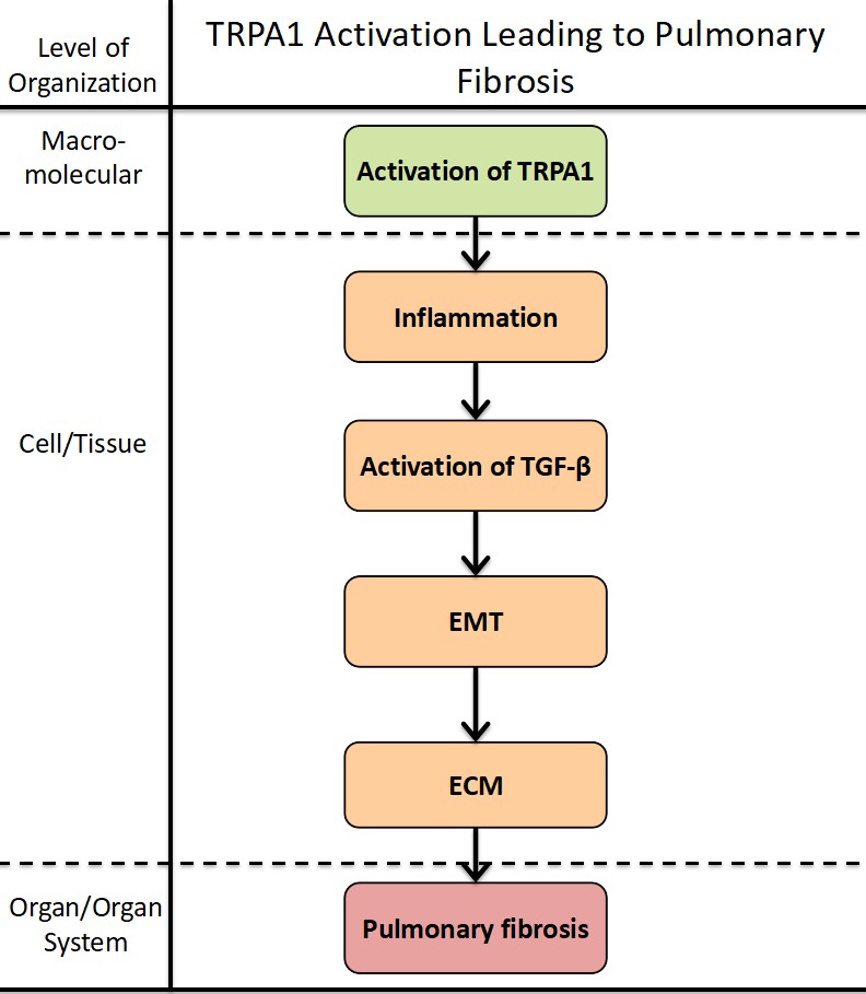
Graphical Representation
Additional AOP Exploration Options
Click links below to explore AOP 655, Activation of Transient Receptor Potential A1 (TRPA1) Leading to Pulmonary Fibrosis in tools offered by third parties.

Point of Contact
Contributors
- Yuan Luo
- Xueyang Lin
Coaches
OECD Information Table
| OECD Project # | OECD Status | Reviewer's Reports | Journal-format Article | OECD iLibrary Published Version |
|---|---|---|---|---|
This AOP was last modified on September 29, 2026 00:44
Revision dates for related pages
| Page | Revision Date/Time |
|---|---|
| TRPA1 activation, TRPA1 Receptor | September 16, 2017 10:17 |
| Increase, Inflammation | February 28, 2024 06:33 |
| Increase, Transforming growth factor-beta signaling | February 11, 2026 05:39 |
| Epithelial Mesenchymal Transition | April 24, 2024 00:44 |
| Increase, Extracellular matrix deposition | February 10, 2026 04:44 |
| Pulmonary fibrosis | May 12, 2023 17:09 |
| TRPA1 activation, TRPA1 Receptor leads to Increase, Inflammation | September 29, 2026 00:42 |
| Increase, Inflammation leads to Activation of TGF-β signaling | September 29, 2026 00:43 |
| Activation of TGF-β signaling leads to EMT | March 18, 2018 09:49 |
| EMT leads to Increase, ECM deposition | September 29, 2026 00:44 |
| Increase, ECM deposition leads to Pulmonary fibrosis | December 06, 2021 16:55 |
Abstract
Abstract: Pulmonary fibrosis is a chronic, irreversible lung disease characterized by excessive extracellular matrix (ECM) deposition and structural destruction of the lungs, closely associated with environmental stressors. Traditional research has primarily focused on the role of classical pathways such as TGF-β/Smad in pulmonary fibrosis, yet the mechanism by which environmental stressors initiate fibrosis remains incompletely elucidated. Transient Receptor Potential Ankyrin 1 (TRPA1), a widely expressed ion channel in lung epithelial cells, fibroblasts, and immune cells, acts as a direct sensor of environmental stress. Its activation triggers downstream calcium signaling and inflammatory responses, but its initiating role in pulmonary fibrosis and association with classical pathways have not been systematically integrated into mechanistic frameworks. Existing OECD AOP Wiki networks also lack a dedicated pathway for TRPA1-mediated pulmonary fibrosis. Therefore, constructing an AOP for TRPA1 activation-induced pulmonary fibrosis is of great significance for revealing novel mechanisms of environmental stressor-induced fibrosis and filling gaps in the existing pulmonary fibrosis AOP network.
AOP Development Strategy
Context
Background: Pulmonary fibrosis is a potentially fatal respiratory disease characterized by the accumulation of interstitial scar tissue, leading to the loss of alveolar function, destruction of normal lung architecture, and impaired respiratory function. Adverse Outcome Pathways (AOP) provide a framework for testing and evaluating pulmonary fibrosis. However, to enhance the applicability of AOPs in regulatory environments, further development of the pulmonary fibrosis AOP framework is necessary. This study develops an AOP for pulmonary fibrosis induced by TRPA1 activation, aimed at predicting all compounds highly related to this AOP. Additionally, this study identifies core Key Events (KEs) shared across different AOPs to facilitate further development of the AOP. Existing OECD AOP Wiki networks also lack a dedicated pathway for TRPA1-mediated pulmonary fibrosis. Therefore, constructing an AOP for TRPA1 activation-induced pulmonary fibrosis is of great significance for revealing novel mechanisms of environmental stressor-induced fibrosis and filling gaps in the existing pulmonary fibrosis AOP network.
Strategy
Summary of the AOP
Events:
Molecular Initiating Events (MIE)
Key Events (KE)
Adverse Outcomes (AO)
| Type | Event ID | Title | Short name |
|---|
| MIE | 1215 | TRPA1 activation, TRPA1 Receptor | TRPA1 activation, TRPA1 Receptor |
| KE | 149 | Increase, Inflammation | Increase, Inflammation |
| KE | 1271 | Increase, Transforming growth factor-beta signaling | Activation of TGF-β signaling |
| KE | 1457 | Epithelial Mesenchymal Transition | EMT |
| KE | 1501 | Increase, Extracellular matrix deposition | Increase, ECM deposition |
| AO | 1458 | Pulmonary fibrosis | Pulmonary fibrosis |
Relationships Between Two Key Events (Including MIEs and AOs)
| Title | Adjacency | Evidence | Quantitative Understanding |
|---|
| TRPA1 activation, TRPA1 Receptor leads to Increase, Inflammation | adjacent | High | High |
| Increase, Inflammation leads to Activation of TGF-β signaling | adjacent | Moderate | Moderate |
| Activation of TGF-β signaling leads to EMT | adjacent | High | High |
| EMT leads to Increase, ECM deposition | adjacent | High | Moderate |
| Increase, ECM deposition leads to Pulmonary fibrosis | adjacent | High | High |
Network View
Prototypical Stressors
Life Stage Applicability
Taxonomic Applicability
Sex Applicability
Overall Assessment of the AOP
Domain of Applicability
Essentiality of the Key Events
|
Type (Event ID) |
Title |
Description |
Support for the essentiality of the KE |
|
MIE (1215) |
Activation of TRPA1 |
The TRPA1 ion channel (also known as the wasabi receptor) is a detector of noxious chemical agents encountered in our environment or produced endogenously during tissue injury or drug metabolism. TRPA1 antagonists hold potential for treating neurogenic inflammatory conditions provoked or exacerbated by irritant exposure1. The TRPA1 channel was first successfully isolated in human lung fibroblasts, and it was found to have a relatively concentrated distribution in the lungs and respiratory tract. It is also involved in various acute and chronic inflammatory processes of lung diseases and may even play a core role in the progression and/or prevention of pulmonary fibrosis2. |
Satyanarayana Achanta and Sven-Eric Jordt (2020)3reported TRPA1 play a crucial role in complex pulmonary pathophysiologic events including, increased intracellular calcium levels, signal transduction, recruitment of proinflammatory cells, neurogenic inflammatory pathways and fibrosis. There is no doubt that TRPA1 localized in the sensory network contribute to airway neurogenic inflammation, and emerging evidence underlines the role of nonneuronal TRPA1 in orchestrating inflammation and repair in the respiratory tract4.
|
|
KE1 (149) |
Increase inflammation |
Inflammatory cytokines refer to a variety of cytokines involved in the inflammatory response. Among the many inflammatory cytokines, TNF-α, IL-1β, IL-6, TGF-β, IL-8, IL-l0, etc5. TNF-α is the earliest and most important inflammatory mediator in the process of inflammatory response, which can activate neutrophils and lymphocytes, increase the permeability of vascular endothelial cells, regulate the metabolic activity of other tissues, and promote the synthesis and release of other cytokines6. IL-6 can induce B-cell differentiation and antibody production, and induce T-cell activation, proliferation and differentiation, and participate in the body's immune response, which is a promoter of inflammatory response7. |
Dysregulated activation of NLRP3 within the pulmonary microenvironment exacerbates inflammation and may incite fibrogenic responses. Contemporary findings suggest that the pro-fibrotic consequences stemming from NLRP3 signaling primarily hinge on the action of interleukin-1β (IL-1β). IL-1β instigates IL-1 receptor signaling, potentiating the activity of transforming growth factor-beta (TGF-β)8. RNA sequencing showed that the tumor necrosis factor alpha (TNFα) signaling pathway and transforming growth factor beta (TGFβ) signaling pathway were consistently activated, potentially contributing to the development of inflammation and fibrosis9. |
|
KE2 (1271) |
Activation of TGF-β signaling |
The transforming growth factor-β (TGF-β) is the prototype of the TGF-β family of growth and differentiation factors, which is encoded by 33 genes in mammals and comprises homo- and heterodimers. This review introduces the reader to the TGF-β family with its complexity of names and biological activities. It also introduces TGF-β as the best-studied factor among the TGF-β family proteins, with its diversity of roles in the control of cell proliferation and differentiation, wound healing and immune system, and its key roles in pathology, for example, skeletal diseases, fibrosis, and cancer10. |
In the presence of persisting injurious pathways, or disrupted repair pathways, activated TGF-β can lead to enhanced epithelial apoptosis and epithelial-to-mesenchymal transition (EMT) as well as fibroblast, and fibrocyte, transformation into myofibroblasts which are resistant to apoptosis11. TGF-β is a central regulator involved in EMT and pulmonary fibrosis. E3 ubiquitin ligases regulate TGF-β-Smad pathway-mediated EMT via the ubiquitin-proteasome pathway12. |
|
KE3 (1457) |
Epithelial mesenchymal transition |
The transdifferentiation of epithelial cells into motile mesenchymal cells, a process known as epithelial-mesenchymal transition (EMT), is integral in development, wound healing and stem cell behaviour, and contributes pathologically to fibrosis and cancer progression. This switch in cell differentiation and behaviour is mediated by key transcription factors, including SNAIL, zinc-finger E-box-binding (ZEB) and basic helix-loop-helix transcription factors, the functions of which are finely regulated at the transcriptional, translational and post-translational levels. The reprogramming of gene expression during EMT, as well as non-transcriptional changes, are initiated and controlled by signalling pathways that respond to extracellular cues. Among these, transforming growth factor-β (TGFβ) family signalling has a predominant role; however, the convergence of signalling pathways is essential for EMT13. |
Using both in vitro and in vivo models, we demonstrated a notable upregulation of epithelial IL5RA during the progression of pulmonary fibrosis. This upregulated IL5RA expression subsequently promotes epithelial-mesenchymal transition (EMT), leading to the generation of mesenchymal phenotype with augmented capability for ECM production14. Epithelial cells can undergo a process called epithelial-mesenchymal transition, in which they acquire a more mesenchymal-like phenotype and contribute directly to fibroblast activation and ECM deposition15. |
|
KE4 (1501) |
Increased extracellular matrix deposition |
Extracellular matrix (ECM) is a non-cellular three-dimensional macromolecular network composed of collagens, proteoglycans/glycosaminoglycans, elastin, fibronectin, laminins, and several other glycoproteins. Matrix components bind each other as well as cell adhesion receptors forming a complex network into which cells reside in all tissues and organs. Cell surface receptors transduce signals into cells from ECM, which regulate diverse cellular functions, such as survival, growth, migration, and differentiation, and are vital for maintaining normal homeostasis. ECM is a highly dynamic structural network that continuously undergoes remodeling mediated by several matrix-degrading enzymes during normal and pathological conditions. Deregulation of ECM composition and structure is associated with the development and progression of several pathologic conditions16. |
Pulmonary fibrosis (PF) is characterized by an increase in collagen synthesis and deposition of extracellular matrix. Several factors, including transforming growth factor-β1 (TGF-β1), mothers against decapentaplegic homolog family proteins (Smad), and alpha-smooth muscle actin (α-SMA) trigger extracellular matrix (ECM) accumulation, fibroblast to myofibroblasts conversion, and epithelial-to-mesenchymal-transition (EMT) leading to PF 17. We demonstrated that the exposure of PM2.5 could enhance the ERS induced-autophagy-mediated Cav-1 degradation, thus activating the TGF-β1/Smad3 axis to promote pneumonocytes apoptosis and overproduction of extracellular matrix (ECM), finally aggravating PF18. |
|
AO (1458) |
Pulmonary fibrosis |
Pulmonary fibrosis is a chronic progressive lung disease that steadily leads to lung architecture disruption and respiratory failure. The development of pulmonary fibrosis is mostly the result of previous acute lung inflammation, caused by a wide variety of etiological factors, not resolved over time and causing the deposition of fibrotic tissue in the lungs19. |
|
Evidence Assessment
|
Upstream event |
Relationship type |
Downstream events |
Evidence from the literature |
|
Activation of TRPA1 |
adjacent |
Increase inflammation |
TRPA1 is expressed in a large subset of sensory nerves, where it integrates numerous noxious stimuli. In non-neuronal cells, TRPA1 also acts as nociceptive sensors and potentiate the inflammatory process20. |
|
|
non-adjacent |
Activation of TGF-β signaling |
The lack of TRPA1 in cultured ocular fibroblasts attenuated expression of TGF-β1, interleukin-6, and α-smooth muscle actin, a myofibroblast the marker, but suppressed the activation of Smad3, p38 MAPK, ERK, and JNK.The responsible mechanism may include the inhibition of TGF-β1-signaling cascades in fibroblasts by attenuated TRPA1 signaling21. |
|
|
non-adjacent |
Epithelial mesenchymal transition |
Transient depotentiation of TRPA1 by siRNA is sufficient to attenuate the downstream signalling pathway of FGFR2c and consequently affect EMT22. |
|
|
non-adjacent |
Increased extracellular matrix deposition |
The activation of transient receptor potential ankyrin 1 (TRPA1) was claimed to be a risk factor in osteoarthritis by causing inflammation and extracellular matrix degradation23. |
|
|
non-adjacent |
Pulmonary fibrosis |
Targeting TRPA1 channels presents promising therapeutic potential in managing pulmonary fibrosis by reducing pro-fibrotic marker expression, inhibiting M2 macrophage polarization, and diminishing collagen deposition24. |
|
Increase inflammation |
adjacent |
Activation of TGF-β signaling |
Dysregulated activation of NLRP3 within the pulmonary microenvironment exacerbates inflammation and may incite fibrogenic responses. Contemporary findings suggest that the pro-fibrotic consequences stemming from NLRP3 signaling primarily hinge on the action of interleukin-1β (IL-1β). IL-1β instigates IL-1 receptor signaling, potentiating the activity of transforming growth factor-beta (TGF-β)8. |
|
|
non-adjacent |
Epithelial mesenchymal transition |
EMT also plays a key role in wound healing processes, which are mediated by inflammatory cells and fibroblasts. Wound healing follows four distinct and successive phases characterized by haemostasis, inflammation, cell proliferation and finally tissue remodeling25. |
|
|
non-adjacent |
Increased extracellular matrix deposition |
The effect of pro- and inflammatory factors on ECM degrading enzymes, such as metalloproteases, and in a more detailed manner on hyaluronan metabolism and the signaling pathways triggered by the binding of hyaluronan with its receptors26. |
|
|
non-adjacent |
Pulmonary fibrosis |
Idiopathic pulmonary fibrosis (IPF) is featured with inflammation and extensive lung remodeling caused by overloaded deposition of extracellular matrix. Scutellarin suppressed BLM-induced inflammation via NF-κB/NLRP3 pathway both in vivo and in vitro. Scutellarin suppressed inflammation and EMT in BLM-induced pulmonary fibrosis through NF-κB/NLRP3 signaling27. |
|
Activation of TGF-β signaling |
adjacent |
Epithelial mesenchymal transition |
it was demonstrated that transforming growth factor (TGF)-beta induces EMT in alveolar epithelial cells (AEC) in vitro and in vivo, and epithelial and mesenchymal markers have been colocalized to hyperplastic type II (AT2) cells in lung tissue from patients with idiopathic pulmonary fibrosis (IPF), suggesting that AEC may exhibit extreme plasticity and serve as a source of fibroblasts and/or myofibroblasts in lung fibrosis28. |
|
|
non-adjacent |
Increased extracellular matrix deposition |
Overexpressed TGF-β causes epithelial-mesenchymal transition (EMT), extracellular matrix (ECM) deposition, cancer-associated fibroblast (CAF) formation, which leads to fibrotic disease, and cancer29. |
|
|
non-adjacent |
Pulmonary fibrosis |
PFD alleviated pulmonary fibrosis in vitro and in vivo through regulating Wnt/GSK-3β/β-catenin and TGF-β1/Smad2/3 signaling pathways, which might further improve the action mechanism of anti-fibrosis effect of PFD30. |
|
Epithelial mesenchymal transition |
adjacent |
Increased extracellular matrix deposition |
In vivo studies revealed that SiNPs-induced pulmonary fibrosis mainly manifested as EMT trans-differentiation in airway epithelial cells, which subsequently led to excessive deposition of extracellular matrix (ECM)31. |
|
|
non-adjacent |
Pulmonary fibrosis |
Epithelial mesenchymal transition (EMT) is a key progression that promotes pulmonary fibrosis (PF). Numb deficiency by siRNA relieved the protection of activating Nrf2 against EMT. Activating Nrf2 antioxidant pathway suppresses EMT during PF via inhibiting the abnormal expression of Numb32. |
|
Increased extracellular matrix deposition |
adjacent |
Pulmonary fibrosis |
It progresses with the recruitment of fibroblasts and myofibroblasts that contribute to the accumulation of extracellular matrix (ECM) proteins, leading to the loss of compliance and alveolar integrity, compromising the gas exchange capacity of the lung33. |
Known Modulating Factors
| Modulating Factor (MF) | Influence or Outcome | KER(s) involved |
|---|---|---|
Quantitative Understanding
Considerations for Potential Applications of the AOP (optional)
References
References
1 Paulsen, C. E., Armache, J. P., Gao, Y., Cheng, Y. & Julius, D. Structure of the TRPA1 ion channel suggests regulatory mechanisms. Nature 520, 511-517, doi:10.1038/nature14367 (2015).
2 Li, C. et al. TRPA1: A promising target for pulmonary fibrosis? Eur J Pharmacol 959, 176088, doi:10.1016/j.ejphar.2023.176088 (2023).
3 Achanta, S. & Jordt, S. E. Transient receptor potential channels in pulmonary chemical injuries and as countermeasure targets. Ann N Y Acad Sci 1480, 73-103, doi:10.1111/nyas.14472 (2020).
4 De Logu, F., Patacchini, R., Fontana, G. & Geppetti, P. TRP functions in the broncho-pulmonary system. Semin Immunopathol 38, 321-329, doi:10.1007/s00281-016-0557-1 (2016).
5 Stepp, M. A. & Menko, A. S. Immune responses to injury and their links to eye disease. Transl Res 236, 52-71, doi:10.1016/j.trsl.2021.05.005 (2021).
6 Jang, D. I. et al. The Role of Tumor Necrosis Factor Alpha (TNF-α) in Autoimmune Disease and Current TNF-α Inhibitors in Therapeutics. Int J Mol Sci 22, doi:10.3390/ijms22052719 (2021).
7 Uciechowski, P. & Dempke, W. C. M. Interleukin-6: A Masterplayer in the Cytokine Network. Oncology 98, 131-137, doi:10.1159/000505099 (2020).
8 Gairola, S., Sinha, A. & Kaundal, R. K. Linking NLRP3 inflammasome and pulmonary fibrosis: mechanistic insights and promising therapeutic avenues. Inflammopharmacology 32, 287-305, doi:10.1007/s10787-023-01389-5 (2024).
9 Hamidi, S. H., Kadamboor Veethil, S. & Hamidi, S. H. Role of pirfenidone in TGF-β pathways and other inflammatory pathways in acute respiratory syndrome coronavirus 2 (SARS-Cov-2) infection: a theoretical perspective. Pharmacol Rep 73, 712-727, doi:10.1007/s43440-021-00255-x (2021).
10 Morikawa, M., Derynck, R. & Miyazono, K. TGF-β and the TGF-β Family: Context-Dependent Roles in Cell and Tissue Physiology. Cold Spring Harb Perspect Biol 8, doi:10.1101/cshperspect.a021873 (2016).
11 Coward, W. R., Saini, G. & Jenkins, G. The pathogenesis of idiopathic pulmonary fibrosis. Ther Adv Respir Dis 4, 367-388, doi:10.1177/1753465810379801 (2010).
12 Inui, N., Sakai, S. & Kitagawa, M. Molecular Pathogenesis of Pulmonary Fibrosis, with Focus on Pathways Related to TGF-β and the Ubiquitin-Proteasome Pathway. Int J Mol Sci 22, doi:10.3390/ijms22116107 (2021).
13 Lamouille, S., Xu, J. & Derynck, R. Molecular mechanisms of epithelial-mesenchymal transition. Nat Rev Mol Cell Biol 15, 178-196, doi:10.1038/nrm3758 (2014).
14 Chen, S., Zhao, T., Xie, S. & Wan, X. Epithelial IL5RA promotes epithelial-mesenchymal transition in pulmonary fibrosis via Jak2/STAT3 cascade. Pulm Pharmacol Ther 84, 102286, doi:10.1016/j.pupt.2024.102286 (2024).
15 Macias-Ceja, D. C., Mendoza-Ballesteros, M. T., Ortega-Albiach, M., Barrachina, M. D. & Ortiz-Masià, D. Role of the epithelial barrier in intestinal fibrosis associated with inflammatory bowel disease: relevance of the epithelial-to mesenchymal transition. Front Cell Dev Biol 11, 1258843, doi:10.3389/fcell.2023.1258843 (2023).
16 Theocharis, A. D., Skandalis, S. S., Gialeli, C. & Karamanos, N. K. Extracellular matrix structure. Adv Drug Deliv Rev 97, 4-27, doi:10.1016/j.addr.2015.11.001 (2016).
17 Cruz, L. C. et al. Identification of tyrosine brominated extracellular matrix proteins in normal and fibrotic lung tissues. Redox Biol 71, 103102, doi:10.1016/j.redox.2024.103102 (2024).
18 Rajesh, R., Atallah, R. & Bärnthaler, T. Dysregulation of metabolic pathways in pulmonary fibrosis. Pharmacol Ther 246, 108436, doi:10.1016/j.pharmthera.2023.108436 (2023).
19 Savin, I. A., Zenkova, M. A. & Sen'kova, A. V. Pulmonary Fibrosis as a Result of Acute Lung Inflammation: Molecular Mechanisms, Relevant In Vivo Models, Prognostic and Therapeutic Approaches. Int J Mol Sci 23, doi:10.3390/ijms232314959 (2022).
20 Gouin, O. et al. TRPV1 and TRPA1 in cutaneous neurogenic and chronic inflammation: pro-inflammatory response induced by their activation and their sensitization. Protein Cell 8, 644-661, doi:10.1007/s13238-017-0395-5 (2017).
21 Okada, Y. et al. TRPA1 is required for TGF-β signaling and its loss blocks inflammatory fibrosis in mouse corneal stroma. Lab Invest 94, 1030-1041, doi:10.1038/labinvest.2014.85 (2014).
22 Mancini, V. et al. TRPA1 Contributes to FGFR2c Signaling and to Its Oncogenic Outcomes in Pancreatic Ductal Adenocarcinoma-Derived Cell Lines. Cancers (Basel) 16, doi:10.3390/cancers16030609 (2024).
23 Che, H. et al. The effect of allyl isothiocyanate on chondrocyte phenotype is matrix stiffness-dependent: Possible involvement of TRPA1 activation. Front Mol Biosci 10, 1112653, doi:10.3389/fmolb.2023.1112653 (2023).
24 Yang, Y. et al. Role of transient receptor potential ankyrin 1 in idiopathic pulmonary fibrosis: modulation of M2 macrophage polarization. Cell Mol Life Sci 81, 187, doi:10.1007/s00018-024-05219-x (2024).
25 Marconi, G. D. et al. Epithelial-Mesenchymal Transition (EMT): The Type-2 EMT in Wound Healing, Tissue Regeneration and Organ Fibrosis. Cells 10, doi:10.3390/cells10071587 (2021).
26 Marozzi, M. et al. Inflammation, Extracellular Matrix Remodeling, and Proteostasis in Tumor Microenvironment. Int J Mol Sci 22, doi:10.3390/ijms22158102 (2021).
27 Peng, L. et al. Scutellarin ameliorates pulmonary fibrosis through inhibiting NF-κB/NLRP3-mediated epithelial-mesenchymal transition and inflammation. Cell Death Dis 11, 978, doi:10.1038/s41419-020-03178-2 (2020).
28 Xiong, R. et al. Histone deacetylase 3 deletion in alveolar type 2 epithelial cells prevents bleomycin-induced pulmonary fibrosis. Clin Epigenetics 15, 182, doi:10.1186/s13148-023-01588-5 (2023).
29 Peng, D., Fu, M., Wang, M., Wei, Y. & Wei, X. Targeting TGF-β signal transduction for fibrosis and cancer therapy. Mol Cancer 21, 104, doi:10.1186/s12943-022-01569-x (2022).
30 Lv, Q. et al. Pirfenidone alleviates pulmonary fibrosis in vitro and in vivo through regulating Wnt/GSK-3β/β-catenin and TGF-β1/Smad2/3 signaling pathways. Mol Med 26, 49, doi:10.1186/s10020-020-00173-3 (2020).
31 Zhou, S. et al. C5a/C5aR1 axis as a key driver promotes epithelial-to-mesenchymal transition in airway epithelial cells in silica nanoparticles-induced pulmonary fibrosis. Int Immunopharmacol 125, 111112, doi:10.1016/j.intimp.2023.111112 (2023).
32 Zhang, Z. et al. Nrf2 antioxidant pathway suppresses Numb-mediated epithelial-mesenchymal transition during pulmonary fibrosis. Cell Death Dis 9, 83, doi:10.1038/s41419-017-0198-x (2018).
33 Zhang, J., Zhang, Y., Chen, Q., Qi, Y. & Zhang, X. The XPO1 inhibitor selinexor ameliorates bleomycin-induced pulmonary fibrosis in mice via GBP5/NLRP3 inflammasome signaling. Int Immunopharmacol 130, 111734, doi:10.1016/j.intimp.2024.111734 (2024).