
The authors have designated this AOP as all rights reserved. Re-use in any form requires advanced permission from the authors.
AOP: 647
Title
ROS formation leading to growth inhibition modulated by the Keap1–Nrf2 antioxidant response
Short name
Graphical Representation
Additional AOP Exploration Options
Click links below to explore AOP 647, ROS formation leading to growth inhibition modulated by the Keap1–Nrf2 antioxidant response in tools offered by third parties.

Point of Contact
Contributors
- Ying Wang
- Li Xie
Coaches
OECD Information Table
| OECD Project # | OECD Status | Reviewer's Reports | Journal-format Article | OECD iLibrary Published Version |
|---|---|---|---|---|
This AOP was last modified on September 02, 2026 16:07
Revision dates for related pages
| Page | Revision Date/Time |
|---|---|
| Increase, Reactive oxygen species | June 12, 2025 01:27 |
| Increase, Cell injury/death | September 02, 2026 04:23 |
| Increase, Oxidative Stress | September 02, 2026 04:19 |
| Increase, Lipid peroxidation | June 23, 2026 06:46 |
| Increase, Protein oxidation | June 23, 2026 06:29 |
| Decrease, Growth | September 02, 2026 03:23 |
| Increase, Cytochrome c release | November 02, 2020 05:57 |
| Increase, Endoplasmic reticulum stress | February 11, 2026 06:00 |
| Activation, caspases | September 14, 2023 08:35 |
| Increase, ROS leads to Increase, Oxidative Stress | June 23, 2026 06:57 |
| Increase, Oxidative Stress leads to Increase, LPO | June 23, 2026 06:59 |
| Increase, Oxidative Stress leads to Increase, Protein oxidation | June 23, 2026 07:04 |
| Increase, LPO leads to Increase, Cyt c release | August 20, 2026 08:05 |
| Increase, Protein oxidation leads to Increase, ER stress | August 20, 2026 08:05 |
| Increase, Cyt c release leads to Caspase activation | September 14, 2023 09:02 |
| Increase, ER stress leads to Caspase activation | August 20, 2026 08:05 |
| Caspase activation leads to Cell injury/death | August 20, 2026 08:06 |
| Cell injury/death leads to Decrease, Growth | June 23, 2026 07:54 |
| Silver nanoparticles | February 15, 2017 03:19 |
Abstract
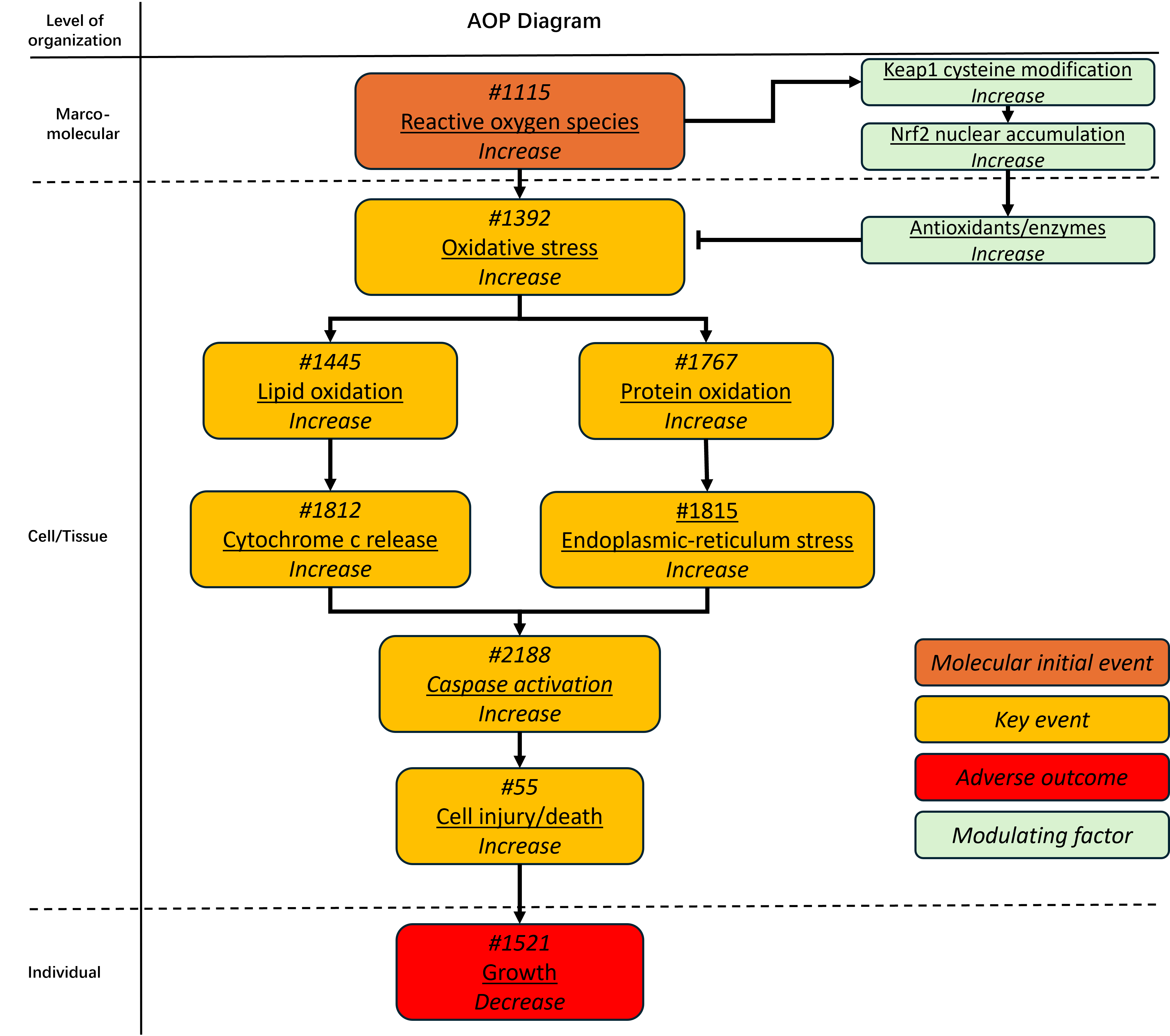
This AOP describes the linkage between increased reactive oxygen species (ROS) formation and decreased organismal growth. The molecular initiating event is increased ROS formation. When ROS production exceeds the buffering capacity of endogenous antioxidant systems, oxidative stress increases. This oxidative burden leads to oxidative damage to cellular macromolecules through two key-event branches: increased lipid oxidation and increased protein oxidation. Increased lipid and protein oxidation can disrupt cellular homeostasis and contribute to mitochondrial and endoplasmic-reticulum stress responses. These processes are represented by increased cytochrome c release and increased endoplasmic-reticulum stress, which promote increased caspase activation. Caspase activation contributes to increased cell injury and cell death. At the organism level, cumulative cellular injury and loss of viable or functional cells can result in decreased growth. The Keap1-Nrf2 antioxidant response is included as a modulating component of this AOP. ROS-mediated modification of Keap1 cysteine residues promotes Nrf2 nuclear accumulation and induction of antioxidant enzymes. This adaptive response can reduce oxidative stress and influence the threshold, magnitude and progression of downstream key events. This AOP provides a mechanistic framework for linking ROS formation, oxidative macromolecular damage, stress-response signalling, apoptotic activation, cell injury and growth impairment. It is considered broadly applicable across aerobic eukaryotes because ROS formation, oxidative damage, antioxidant defence and regulated cell-death processes are conserved biological features. Growth impairment is included as the adverse outcome because it is a biologically and ecotoxicologically relevant endpoint across multiple taxa.
AOP Development Strategy
Context
Reactive oxygen species (ROS) are produced during normal aerobic metabolism and act as physiological signalling molecules. Their biological effects depend on concentration, duration, cellular location and antioxidant capacity. At controlled levels, ROS participate in redox regulation; when ROS production exceeds antioxidant and repair capacity, oxidative stress can disrupt redox signalling and damage cellular macromolecules (Sies and Jones, 2020; Sies, 2015). In aquatic animals, environmentally induced oxidative stress is a common response to chemical and non-chemical stressors, including metals, pesticides, nanoparticles, radiation and changes in environmental conditions (Lushchak, 2011; Song et al., 2023).
Cells maintain redox homeostasis through antioxidant enzymes, low-molecular-weight redox buffers and stress-response pathways. The Keap1-Nrf2 pathway is a major adaptive defence system against oxidative and electrophilic stress. Keap1 represses Nrf2-dependent activation of antioxidant response elements under basal conditions, while oxidative or electrophilic modification of Keap1 promotes Nrf2 activation and induction of antioxidant and detoxification genes (Itoh et al., 1999; Baird and Yamamoto, 2020). This adaptive response is important for interpreting oxidative-stress outcomes because it can delay, reduce or modify downstream toxicity depending on exposure intensity, duration and biological capacity for compensation. Sustained oxidative stress can affect cell fate through damage to lipids, proteins and other macromolecules, and through interactions with mitochondrial and endoplasmic-reticulum (ER) stress responses. Cytochrome c has a central role in mitochondrial apoptosis, including cardiolipin oxidation and release of proapoptotic factors, followed by Apaf-1/caspase-9 complex formation and activation of the apoptotic protease cascade (Kagan et al., 2005; Li et al., 1997). ER stress is also linked to cell-fate decisions and can contribute to apoptosis when adaptive unfolded-protein responses are insufficient (Cao and Kaufman, 2014; Chen et al., 2023).
Growth impairment is a regulatory and ecotoxicologically relevant apical outcome because growth integrates energy acquisition, metabolism, cellular proliferation, tissue maintenance and development over time. Previous work on AOP development for chronic toxicity has used growth impairment to illustrate how mechanistic information can support interpretation of adverse outcomes relevant to hazard assessment (Groh et al., 2015). The present AOP was developed to organise conserved oxidative-stress biology in a form useful for mechanistic interpretation, chemical prioritisation and future integrated approaches to testing and assessment.
Strategy
Summary of the AOP
Events:
Molecular Initiating Events (MIE)
Key Events (KE)
Adverse Outcomes (AO)
| Type | Event ID | Title | Short name |
|---|
| MIE | 1115 | Increase, Reactive oxygen species | Increase, ROS |
| KE | 1392 | Increase, Oxidative Stress | Increase, Oxidative Stress |
| KE | 1445 | Increase, Lipid peroxidation | Increase, LPO |
| KE | 1767 | Increase, Protein oxidation | Increase, Protein oxidation |
| KE | 1812 | Increase, Cytochrome c release | Increase, Cyt c release |
| KE | 1815 | Increase, Endoplasmic reticulum stress | Increase, ER stress |
| KE | 2188 | Activation, caspases | Caspase activation |
| KE | 55 | Increase, Cell injury/death | Cell injury/death |
| AO | 1521 | Decrease, Growth | Decrease, Growth |
Relationships Between Two Key Events (Including MIEs and AOs)
| Title | Adjacency | Evidence | Quantitative Understanding |
|---|
| Increase, ROS leads to Increase, Oxidative Stress | adjacent | High | High |
| Increase, Oxidative Stress leads to Increase, LPO | adjacent | High | High |
| Increase, Oxidative Stress leads to Increase, Protein oxidation | adjacent | High | Moderate |
| Increase, LPO leads to Increase, Cyt c release | adjacent | High | Low |
| Increase, Protein oxidation leads to Increase, ER stress | adjacent | High | Low |
| Increase, Cyt c release leads to Caspase activation | adjacent | High | Low |
| Increase, ER stress leads to Caspase activation | adjacent | High | Low |
| Caspase activation leads to Cell injury/death | adjacent | High | Low |
| Cell injury/death leads to Decrease, Growth | adjacent | High | High |
Network View
Prototypical Stressors
| Name |
|---|
| Silver nanoparticles |
Life Stage Applicability
| Life stage | Evidence |
|---|---|
| Not Otherwise Specified | High |
Taxonomic Applicability
Sex Applicability
| Sex | Evidence |
|---|---|
| Unspecific | High |
Overall Assessment of the AOP
Domain of Applicability
Essentiality of the Key Events
Evidence Assessment
Known Modulating Factors
| Modulating Factor (MF) | Influence or Outcome | KER(s) involved |
|---|---|---|
Quantitative Understanding
Considerations for Potential Applications of the AOP (optional)
References
Chen, X., Shi, C., He, M., Xiong, S., & Xia, X. (2023). Endoplasmic reticulum stress: Molecular mechanism and therapeutic targets. Signal Transduction and Targeted Therapy, 8, 352. https://doi.org/10.1038/s41392-023-01570-w
Groh, K. J., Carvalho, R. N., Chipman, J. K., Denslow, N. D., Halder, M., Murphy, C. A., Roelofs, D., Rolaki, A., Schirmer, K., & Watanabe, K. H. (2015). Development and application of the adverse outcome pathway framework for understanding and predicting chronic toxicity: II. A focus on growth impairment in fish. Chemosphere, 120, 778-792. https://doi.org/10.1016/j.chemosphere.2014.10.006
Itoh, K., Wakabayashi, N., Katoh, Y., Ishii, T., Igarashi, K., Engel, J. D., & Yamamoto, M. (1999). Keap1 represses nuclear activation of antioxidant responsive elements by Nrf2 through binding to the amino-terminal Neh2 domain. Genes & Development, 13(1), 76-86. https://doi.org/10.1101/gad.13.1.76
Kagan, V. E., Tyurin, V. A., Jiang, J., Tyurina, Y. Y., Ritov, V. B., Amoscato, A. A., Osipov, A. N., Belikova, N. A., Kapralov, A. A., Kini, V., Vlasova, I. I., Zhao, Q., Zou, M., Di, P., Svistunenko, D. A., Kurnikov, I. V., & Borisenko, G. G. (2005). Cytochrome c acts as a cardiolipin oxygenase required for release of proapoptotic factors. Nature Chemical Biology, 1(4), 223-232. https://doi.org/10.1038/nchembio727
Li, P., Nijhawan, D., Budihardjo, I., Srinivasula, S. M., Ahmad, M., Alnemri, E. S., & Wang, X. (1997). Cytochrome c and dATP-dependent formation of Apaf-1/caspase-9 complex initiates an apoptotic protease cascade. Cell, 91(4), 479-489. https://doi.org/10.1016/S0092-8674(00)80434-1
Lushchak, V. I. (2011). Environmentally induced oxidative stress in aquatic animals. Aquatic Toxicology, 101(1), 13-30. https://doi.org/10.1016/j.aquatox.2010.10.006
Sies, H. (2015). Oxidative stress: A concept in redox biology and medicine. Redox Biology, 4, 180-183. https://doi.org/10.1016/j.redox.2015.01.002
Sies, H., & Jones, D. P. (2020). Reactive oxygen species (ROS) as pleiotropic physiological signalling agents. Nature Reviews Molecular Cell Biology, 21(7), 363-383. https://doi.org/10.1038/s41580-020-0230-3
Song, C., Sun, C., Liu, B., & Xu, P. (2023). Oxidative stress in aquatic organisms. Antioxidants, 12(6), 1223. https://doi.org/10.3390/antiox12061223