AOP ID and Title:
Graphical Representation
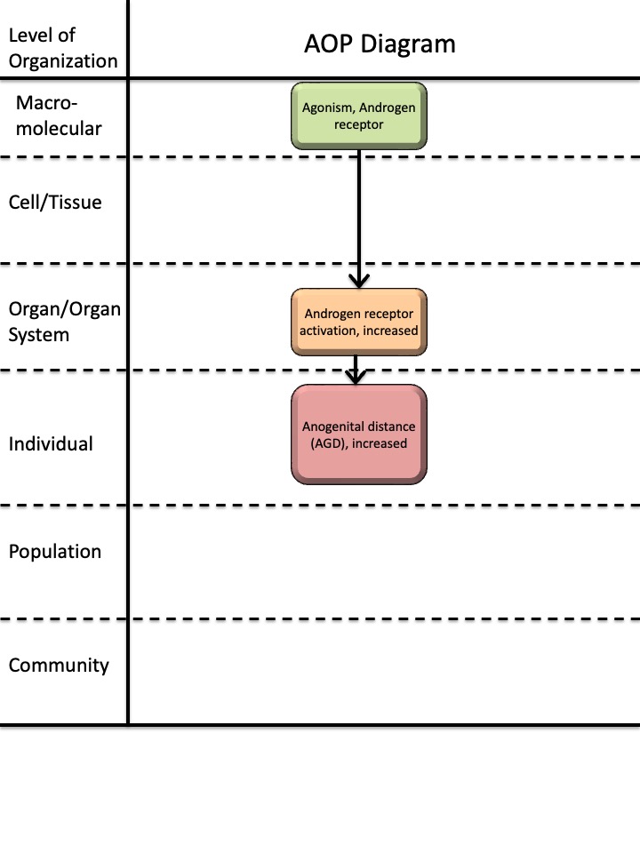
Status
| Author status | OECD status | OECD project | SAAOP status |
|---|---|---|---|
| Under development: Not open for comment. Do not cite |
Abstract
The purpose of developing this AOP was to establish a framework for identifying endocrine disruptors based on the measurement of anogenital distance (AGD) in female rodents. AGD is defined as the distance between the anus and the external genitalia. In both rodents and humans, female AGD is approximately half the length of male AGD (Liu et al., 2014; Salazar-Martinez et al., 2004; Schwartz et al., 2019; Sharpe, 2020; Thankamony et al., 2016; Wise, 2024). This difference reflects androgen-dependent regulation of perineal growth during fetal development in males, whereas in females, the absence of androgens limits this growth (Ipulan et al., 2016; Schwartz et al., 2019; Sharpe, 2020; Welsh et al., 2008).
AGD is considered an apical endpoint and is included in OECD test guidelines TG414, TG416, TG421, TG422 and TG443 (OECD, 2001; OECD, 2018; OECD, 2025a; OECD, 2025b; OECD, 2025c). According to ECHA/EFSA guidance for identifying endocrine disruptors, AGD is classified as an EATS-mediated parameter and is used in the identification of endocrine disruptors (ECHA/EFSA, 2018).
The molecular initiating event (MIE) for this AOP is androgen receptor (AR) agonism, meaning the binding of a compound to the AR that activates the receptor. This leads to the first key event (KE): increased AR activation in tissues and organs in vivo, including the perineum. During fetal development in females, increased AR activation in the perineum results in increased AGD considered an adverse outcome (AO).
The overall weight of evidence supporting this AOP is high. Biological plausibility for both key event relationships (KERs) is strong, as androgen receptor mechanisms and androgenic effects on AGD are well established. Empirical evidence for the KERs is based on studies showing that AR agonists increase female AGD in prenatal rat studies.
This AOP is applicable for identifying androgenic endocrine disruptors.
AOP Development Strategy
Context
This AOP was developed as part of the European Food Safety Authority (EFSA) project (Grant Agreement No. GP/EFSA/PREV/2022/01) aimed at creating AOPs for endocrine disruptors. The purpose of this AOP is to provide a framework for identifying endocrine disruptors associated with increased AGD in female rodent offspring. Other AOPs within the project address shortened AGD in males (AOPs 305, 306, and 307).
The molecular initiating event (MIE25: Agonism, Androgen receptor) and key event (KE2274: Androgen receptor activation, increased) described in this AOP can serve as a foundation for developing additional AOPs related to adverse outcomes resulting from androgen receptor agonism.
Furthermore, this AOP can be expanded into an AOP network by incorporating pathways such as “Increased testosterone levels leading to long anogenital distance in female offspring” and “Increased dihydrotestosterone levels leading to long anogenital distance in female offspring.” These can be developed using the key events (KE2272, KE2273) and key event relationships (KER3380, KER3381) established in the EFSA-funded project.
A shortened AGD is recognized as a sensitive marker of reduced AR activity during the masculinization programming window in the male, whereas in the female a longer AGD is a marker of increased AR activity and masculinization of the female fetus in this period.
Strategy
This AOP was developed in two steps.
First, an upstream AOP network for “increased androgen activity” was constructed. The AOP-Wiki was screened to identify existing key events (KEs) related to increased androgen activity by searching KE and KER pages for the terms androgen, testosterone, dihydrotestosterone, and DHT. Two relevant KEs were identified, but no relevant KERs. KE25 was updated, while KE286 required no revision. All other KEs and KERs were newly developed following the methodology outlined in the AOP Developers’ Handbook (version 2.7): KE2272, KE2273, KE2274, KER3378, KER3379, KER3380, and KER3381.
The network reflects processes widely regarded as canonical, and evidence was primarily drawn from review articles and book chapters identified through searches in PubMed and library catalogues. For the current AOP, the following KEs from the upstream network were used: KE25, KE2274, and KER3378. The remaining KEs and KERs could be used to expand the current AOP into an AOP network as described above.
In the second step, KE2365 and KER3628 were developed.
Evidence for KE2365 was drawn mainly from review articles identified through PubMed searches. A systematic weight-of-evidence approach was applied to collect, evaluate, extract, and integrate evidence for KER3628, as detailed on its dedicated page. Briefly, a scoping literature search was first conducted in PubMed to identify relevant model substances. Based on these findings, a targeted search was performed in PubMed and Web of Science using terms related to the model substances and AGD.
Study reliability for included publications was assessed using the Science in Risk Assessment and Policy (SciRAP) tools for in vivo studies (http://www.scirap.org), and overall confidence in the evidence for each model substance was categorized.
The AOP includes three KEs: the MIE, the AO, and one intermediate KE.
These were selected to capture the main steps in the pathway from AR agonism to increased AGD. Additional KEs could be added, such as KE286 (Altered transcription of genes by the AR) and a KE describing androgen regulation of perineal growth, which likely involves androgen-mediated development of the levator ani and bulbocavernosus (LABC) muscle complex in the perineum. However, sufficient empirical evidence for these related KERs is currently lacking.
Summary of the AOP
Events
Molecular Initiating Events (MIE), Key Events (KE), Adverse Outcomes (AO)
| Sequence | Type | Event ID | Title | Short name |
|---|---|---|---|---|
| MIE | 25 | Agonism, Androgen receptor | Agonism, Androgen receptor | |
| KE | 2274 | Androgen receptor activation, increased | Androgen receptor activation, increased | |
| AO | 2365 | Anogenital distance (AGD), increased | Anogenital distance (AGD), increased |
Key Event Relationships
| Upstream Event | Relationship Type | Downstream Event | Evidence | Quantitative Understanding |
|---|---|---|---|---|
| Agonism, Androgen receptor | adjacent | Androgen receptor activation, increased | High | Low |
| Androgen receptor activation, increased | adjacent | Anogenital distance (AGD), increased | High | Low |
Stressors
| Name | Evidence |
|---|---|
| 17-Methyltestosterone | |
| Testosterone propionate | |
| 17beta-Trenbolone |
Overall Assessment of the AOP
Domain of Applicability
Life Stage Applicability| Life Stage | Evidence |
|---|---|
| Fetal | High |
| Sex | Evidence |
|---|---|
| Female | High |
Life stage applicability
The life stage applicability domain for this AOP is fetal. Although AR activation occurs during fetal development, puberty, and adulthood, the regulation of AGD growth is specifically controlled by androgens during the masculinization programming window, which takes place in fetal development (Dalton & Gao, 2010; Luetjens & Weinbauer, 2012; Naamneh Elzenaty et al., 2022; Sutinen et al., 2017; Welsh et al., 2008).
Taxonomic applicability
The biologically plausible taxonomic applicability domain for this AOP is mammals, as fetal masculinization is regulated by androgens across all mammalian species (Welsh et al., 2014). The empirical taxonomic applicability domain is rat and human, supported by prenatal studies in rat demonstrating that androgen receptor activation leads to a measurable increase in female offspring AGD (Armoskus et al., 2014; Guerra et al., 2014; Hotchkiss et al., 2007; Sathishkumar et al., 2011; Welsh et al., 2009; Wilson et al., 2002; Wolf et al., 2002; Wolf et al., 2004) and epidemiological studies suggesting that elevated androgen levels in women are associated with increased AGD (Mira-Escolano et al., 2014; Pan et al., 2021; Zamani et al., 2023).
Sex applicability
The sex applicability domain for this AOP is females, supported by studies showing effects of androgen activation on female AGD (Armoskus et al., 2014; Guerra et al., 2014; Hotchkiss et al., 2007; Mira-Escolano et al., 2014; Pan et al., 2021; Sathishkumar et al., 2011; Welsh et al., 2009; Wilson et al., 2002; Wolf et al., 2002; Wolf et al., 2004; Zamani et al., 2023).
Essentiality of the Key Events
|
KE |
Evidence |
Level of evidence |
|
MIE25 |
Direct evidence on effect on KE2274: Studies in AR knockout mice demonstrate that AR is essential for mediating effects on the reproductive system by AR activation (De Gendt et al., 2004; Holdcraft & Braun, 2004; Matsumoto et al., 2003; Notini et al., 2005; Yeh et al., 2002). AR antagonists flutamide, procymidone and vinclozolin inhibit AR activity in vivo as shown in the OECD test guideline Hershberger assay (Browne et al., 2018). Direct evidence on effect on AO2365: Studies in AR knockout mice demonstrate that AR is essential for a longer AGD, as male AR knockout mice exhibit AGD lengths comparable to female wild-type mice (Yeh et al., 2002, Sato et al., 2004). It should be noted AGD in male mice was studied but the AO2365 is in females. Prenatal exposure to the AR antagonists flutamide, procymidone and vinclozolin results in shorter AGD in rat studies (Casto et al., 2003; Foster & Harris, 2005; Fussell et al., 2015; Goto et al., 2004; Gray et al., 1994; Hass et al., 2007; Hass et al., 2012; Inawaka et al., 2010; Kita et al., 2016; Ostby et al., 1999; McIntyre et al., 2001; Shimamura et al., 2002; Wolf et al., 2000; Yamasaki et al., 2005). It should be noted AGD in male rats was studied but the AO2365 is in females. |
High. There is direct evidence from knockout and antagonist studies. There are no inconsistencies identified but it should be noted that the effect on AGD was studied in male animals. |
|
KE2274 |
Direct evidence on effect on AO2365: Studies in AR knockout mice demonstrate that AR activity is essential for a longer AGD, as male AR knockout mice exhibit AGD lengths comparable to female wild-type mice (Yeh et al., 2002, Sato et al., 2004). It should be noted AGD in male mice was studied but the AO2365 is in females. Prenatal exposure to the AR antagonists flutamide, procymidone and vinclozolin results in reduced AGD in rat studies (Casto et al., 2003; Foster & Harris, 2005; Fussell et al., 2015; Goto et al., 2004; Gray et al., 1994; Hass et al., 2007; Hass et al., 2012; Inawaka et al., 2010; Kita et al., 2016; Ostby et al., 1999; McIntyre et al., 2001; Shimamura et al., 2002; Wolf et al., 2000; Yamasaki et al., 2005). It should be noted AGD in male rats was studied but the AO2365 is in females. |
High. There is direct evidence from knockout and antagonist studies. There are no inconsistencies identified, but it should be noted that the effect on AGD was studied in male animals. |
Weight of Evidence Summary
Assessment of biological plausibility
|
KER |
Assessment |
|
KER 3378: Agonism, Androgen receptor leads to Androgen receptor activation, increased |
The biological plausibility of the KER is High. It is a generally recognised process, canonical knowledge, that binding of agonists to AR leads to increased AR activation in vivo (Dalton & Gao, 2010; Luetjens & Weinbauer, 2012; Naamneh Elzenaty et al., 2022; Sutinen et al., 2017). |
|
KER 3628: Androgen receptor activation, increased leads to AGD, increased |
The biological plausibility of the KER is High. It is a well-documented by numerous studies in rats that AR activation drives the fetal masculinization process, including the growth of the perineum, which can be measured as AGD (Liu et al., 2014; Salazar-Martinez et al, 2004; Schwartz et al., 2019; Sharpe, 2020; Thankamony et al., 2016; Welsh et al., 2008; Wise, 2024). This is supported by epidemiological studies (Mira-Escolano et al., 2014; Pan et al., 2021; Zamani et al., 2023). |
Assessment of empirical evidence
|
KER |
Assessment |
|
KER 3378: Agonism, Androgen receptor leads to Androgen receptor activation, increased |
The empirical support for the KER is High. AR agonists such as testosterone propionate, methyl testosterone and trenbolone have been identified as agonists in the US EPA Androgen receptor pathway model, which integrates in vitro assays for receptor binding, coregulator recruitment and transactivation. These stressors have also been shown to increase androgen activity in Hershberger assay and other in vivo assays (Kleinstreuer et al., 2018; Browne et al., 2018). Assessing dose and temporal concordance for this KER is challenging because the upstream KE is measured in vitro, whereas the downstream KE reflects on increased in vivo activation. There are no available data on incidence concordance. |
|
KER 3628: Androgen receptor activation, increased leads to AGD, increased |
The empirical support for the KER is High. The empirical evidence is based on four model substances: methyl testosterone, testosterone, testosterone propionate and trenbolone. These substances bind AR and act as AR agonists, as demonstrated in in vitro studies (Kleinstreuer et al., 2018; Judson et al., 2020) and androgen activity in in vivo Hershberger assays (Browne et al., 2018). Prenatal exposure to these substances in rat studies has been shown to increase AGD in female offspring, as reported in multiple studies (Armoskus et al., 2014; Guerra et al., 2014; Hotchkiss et al., 2007; Juarez et al., 1995; Kawashima et al., 1975; McCoy et al., 1992; Rhees et al., 1997; Tehrani et al., 2014; Welsh et al., 2009; Wilson et al., 2002; Wolf et al., 2002; Wolf et al., 2004; Wu et al., 2010) Assessing dose and temporal concordance for this KER is challenging because the upstream KE is measured in vitro, whereas the downstream KE is assessed in vivo. The upstream KE, increased AR activation, is expected to occur rapidly, within minutes to hours after exposure to the substance (Naamneh Elzenaty et al., 2022; Sutinen et al., 2017). In the prenatal in vivo studies, the exposure occurred during GD15–19, with AGD measured in female offspring between GD21 and PND30. No evidence is currently available to assess incidence concordance for this KER. |
Quantitative Consideration
The level of quantitative understanding of the AOP is low. No specific evidence has been identified for response-response relationships for the KERs or for the presence of feedforward or feedback loops. There is some understanding of the time-scale. Agonism of the AR leads rapidly to increased AR activity in vivo. Effects on activation of the receptor on cellular function can be seen after minutes to hours (Naamneh Elzenaty et al., 2022; Sutinen et al., 2017). In prenatal in vivo studies, exposure to a substance that activates AR for five to six days during the masculinization window (around GD16-20 in rats) can result in increased AGD at PND2 (Hotchkiss et al., 2007; Wilson et al., 2002; Wolf et al., 2004).
Considerations for Potential Applications of the AOP (optional)
AGD is considered an apical endpoint and is included in OECD test guidelines TG414, TG416, TG421, TG422, and TG443 (OECD, 2001; OECD, 2018; OECD, 2025a; OECD, 2025b; OECD, 2025c). According to ECHA/EFSA guidance for identifying endocrine disruptors, AGD is classified as an EATS-mediated parameter and is used in the identification of endocrine-disrupting substances (ECHA/EFSA, 2018).
This AOP supports the regulatory use of increased AGD in female rodents as an indicator of endocrine disruption, specifically to identify androgenic endocrine disruptors.
References
Armoskus, C., Mota, T., Moreira, D., & Tsai, H.-W. (2014). Effects of Prenatal Testosterone Exposure on Sexually Dimorphic Gene Expression in the Neonatal Mouse Cortex and Hippocampus. Journal of Steroids & Hormonal Science, 5(3), 1000139.
Browne, P., Kleinstreuer, N. C., Ceger, P., Deisenroth, C., Baker, N., Markey, K., Thomas, R. S., Judson, R. J., & Casey, W. (2018). Development of a curated Hershberger database. Reproductive Toxicology, 81, 259–271. https://doi.org/10.1016/j.reprotox.2018.08.016
Casto, J., Ward, O., & Bartke, A. (2003). Play, copulation, anatomy, and testosterone in gonadally intact male rats prenatally exposed to flutamide. Physiology & Behavior, 79(4), 633–641. https://doi.org/10.1016/S0031-9384(03)00120-3
Dalton, J. T., & Gao, W. (2010). Androgen Receptor. In C. M. Bunce & M. J. Campbell (Eds), Nuclear Receptors (pp. 143–182). Springer Netherlands. https://doi.org/10.1007/978-90-481-3303-1_6
De Gendt, K., Swinnen, J. V., Saunders, P. T. K., Schoonjans, L., Dewerchin, M., Devos, A., Tan, K., Atanassova, N., Claessens, F., Lécureuil, C., Heyns, W., Carmeliet, P., Guillou, F., Sharpe, R. M., & Verhoeven, G. (2004). A Sertoli cell-selective knockout of the androgen receptor causes spermatogenic arrest in meiosis. Proceedings of the National Academy of Sciences, 101(5), 1327–1332. https://doi.org/10.1073/pnas.0308114100
European Chemical Agency (ECHA) and European Food Safety Authority (EFSA) with the technical support of the Joint Research Centre (JRC), Andersson, N., Arena, M., Auteri, D., Barmaz, S., Grignard, E., Kienzler, A., Lepper, P., Lostia, A. M., Munn, S., Parra Morte, J. M., Pellizzato, F., Tarazona, J., Terron, A., & Van der Linden, S. (2018). Guidance for the identification of endocrine disruptors in the context of Regulations (EU) No 528/2012 and (EC) No 1107/2009. EFSA Journal, 16(6). https://doi.org/10.2903/j.efsa.2018.5311
Foster PM & Harris MW. (2005). Changes in androgen-mediated reproductive development in male rat offspring following exposure to a single oral dose of flutamide at different gestational ages. Toxicological Sciences : An Official Journal of the Society of Toxicology, 85(2), 1024–1032. https://doi.org/10.1093/toxsci/kfi159
Fussell KC, Schneider S, Buesen R, Groeters S, Strauss V, Melching-Kollmuss S, & van Ravenzwaay B. (2015). Investigations of putative reproductive toxicity of low-dose exposures to flutamide in Wistar rats. Archives of Toxicology, 89(12), 2385–2402. https://doi.org/10.1007/s00204-015-1622-6
Goto K, Koizumi K, Takaori H, Fujii Y, Furuyama Y, Saika O, Suzuki H, Saito K, & Suzuki K. (2004). Effects of flutamide on sex maturation and behavior of offspring born to female rats treated during late pregnancy. The Journal of Toxicological Sciences, 29(5), 517–534. https://doi.org/10.2131/jts.29.517
Gray LE Jr, Ostby JS, & Kelce WR. (1994). Developmental effects of an environmental antiandrogen: The fungicide vinclozolin alters sex differentiation of the male rat. Toxicology and Applied Pharmacology, 129(1), 46–52. https://doi.org/10.1006/taap.1994.1227
Guerra, M., Silva, R., Luchiari, H., Sanabria, M., & Kempinas, W. (2014). Perinatal Androgenic Exposure and Reproductive Health Effects Female Rat Offspring. Journal of Toxicology and Environmental Health-Part A-Current Issues, 77(7), 375–389. https://doi.org/10.1080/15287394.2013.874881
Hass, U., Boberg, J., Christiansen, S., Jacobsen, P. R., Vinggaard, A. M., Taxvig, C., Poulsen, M. E., Herrmann, S. S., Jensen, B. H., Petersen, A., Clemmensen, L. H., & Axelstad, M. (2012). Adverse effects on sexual development in rat offspring after low dose exposure to a mixture of endocrine disrupting pesticides. Reproductive Toxicology, 34(2), 261–274. https://doi.org/10.1016/j.reprotox.2012.05.090
Hass U, Scholze M, Christiansen S, Dalgaard M, Vinggaard AM, Axelstad M, Metzdorff SB, & Kortenkamp A. (2007). Combined exposure to anti-androgens exacerbates disruption of sexual differentiation in the rat. Environmental Health Perspectives, 115, 122–128. https://doi.org/10.1289/ehp.9360
Holdcraft, R. W., & Braun, R. E. (2004). Androgen receptor function is required in Sertoli cells for the terminal differentiation of haploid spermatids. Development, 131(2), 459–467. https://doi.org/10.1242/dev.00957
Hotchkiss, A. K., Lambright, C. S., Ostby, J. S., Parks-Saldutti, L., Vandenbergh, J. G., & Gray, L. E. J. (2007). Prenatal testosterone exposure permanently masculinizes anogenital distance, nipple development, and reproductive tract morphology in female Sprague-Dawley rats. Toxicological Sciences : An Official Journal of the Society of Toxicology, 96(2), 335–345. https://doi.org/10.1093/toxsci/kfm002
Inawaka, K., Kishimoto, N., Higuchi, H., & Kawamura, S. (2010). Maternal exposure to procymidone has no effects on fetal external genitalia development in male rabbit fetuses in a modified developmental toxicity study. Journal of Toxicological Sciences, 35(3), 299–307. https://doi.org/10.2131/jts.35.299
Ipulan, L. A., Raga, D., Suzuki, K., Murashima, A., Matsumaru, D., Cunha, G., & Yamada, G. (2016). Investigation of sexual dimorphisms through mouse models and hormone/hormone-disruptor treatments. Differentiation; Research in Biological Diversity, 91(4–5), 78–89. https://doi.org/10.1016/j.diff.2015.11.001
Juárez, J., Corsi-Cabrera, M., & del Río-Portilla, I. (1995). Effects of prenatal testosterone treatment on sex differences in the EEG activity of the rat. Brain Research, 694(1–2), 21–28. https://doi.org/10.1016/0006-8993(95)00725-6
Judson, R., Houck, K., Paul Friedman, K., Brown, J., Browne, P., Johnston, P. A., Close, D. A., Mansouri, K., & Kleinstreuer, N. (2020). Selecting a minimal set of androgen receptor assays for screening chemicals. Regulatory Toxicology and Pharmacology, 117, 104764. https://doi.org/10.1016/j.yrtph.2020.104764
Kawashima, K., Nakaura, S., Nagao, S., Tanaka, S., & Kuwamura, T. (1975). Quantitative evaluation of virilizing activity of steroids by measuring morphological changes in uro-genital region of rats. Endocrinologia Japonica, 22(5), 439–444. https://doi.org/10.1507/endocrj1954.22.439
Kita DH, Meyer KB, Venturelli AC, Adams R, Machado DL, Morais RN, Swan SH, Gennings C, & Martino-Andrade AJ. (2016). Manipulation of pre and postnatal androgen environments and anogenital distance in rats. Toxicology, 368, 152–161. https://doi.org/10.1016/j.tox.2016.08.021
Kleinstreuer, N. C., Browne, P., Chang, X., Judson, R., Casey, W., Ceger, P., Deisenroth, C., Baker, N., Markey, K., & Thomas, R. S. (2018). Evaluation of androgen assay results using a curated Hershberger database. Reproductive Toxicology, 81, 272–280. https://doi.org/10.1016/j.reprotox.2018.08.017
Liu, C., Xu, X., & Huo, X. (2014). Anogenital distance and its application in environmental health research. Environmental Science and Pollution Research International, 21(8), 5457–5464. https://doi.org/10.1007/s11356-014-2570-z
Luetjens, C. M., & Weinbauer, G. F. (2012). Testosterone: Biosynthesis, transport, metabolism and (non-genomic) actions. In E. Nieschlag, H. M. Behre, & S. Nieschlag (Eds), Testosterone (4th edn, pp. 15–32). Cambridge University Press. https://doi.org/10.1017/CBO9781139003353.003
Matsumoto, T., Takeyama, K., Sato, T., & Kato, S. (2003). Androgen receptor functions from reverse genetic models. The Journal of Steroid Biochemistry and Molecular Biology, 85(2–5), 95–99. https://doi.org/10.1016/s0960-0760(03)00231-0
McCoy, S. J., & Shirley, B. A. (1992). Effects of prenatal administration of testosterone and cortisone on the reproductive system of the female rat. Life Sciences, 50(9), 621–628. https://doi.org/10.1016/0024-3205(92)90248-n
McIntyre BS, Barlow NJ, & Foster PM. (2001). Androgen-mediated development in male rat offspring exposed to flutamide in utero: Permanence and correlation of early postnatal changes in anogenital distance and nipple retention with malformations in androgen-dependent tissues. Toxicological Sciences : An Official Journal of the Society of Toxicology, 62(2), 236–249. (rayyan-423946802). https://doi.org/10.1093/toxsci/62.2.236
Mira-Escolano, M., Mendiola, J., Mínguez-Alarcón, L., Roca, M., Cutillas-Tolín, A., López-Espín, J., & Torres-Cantero, A. (2014). Anogenital distance of women in relation to their mother’s gynaecological characteristics before or during pregnancy. Reproductive Biomedicine Online, 28(2), 209–215. https://doi.org/10.1016/j.rbmo.2013.09.026
Naamneh Elzenaty, R., Du Toit, T., & Flück, C. E. (2022). Basics of androgen synthesis and action. Best Practice & Research Clinical Endocrinology & Metabolism, 36(4), 101665. https://doi.org/10.1016/j.beem.2022.101665
Notini, A. J., Davey, R. A., McManus, J. F., Bate, K. L., & Zajac, J. D. (2005). Genomic actions of the androgen receptor are required for normal male sexual differentiation in a mouse model. Journal of Molecular Endocrinology, 35(3), 547–555. https://doi.org/10.1677/jme.1.01884
OECD. (2001). Test No. 416: Two-Generation Reproduction Toxicity. OECD. https://doi.org/10.1787/9789264070868-en
OECD. (2018). Test No. 414: Prenatal Developmental Toxicity Study. OECD. https://doi.org/10.1787/9789264070820-en
OECD. (2025a). Test No. 421: Reproduction/Developmental Toxicity Screening Test. OECD Publishing. https://doi.org/10.1787/9789264264380-en
OECD. (2025b). Test No. 422: Combined Repeated Dose Toxicity Study with the Reproduction/Developmental Toxicity Screening Test. OECD Publishing. https://doi.org/10.1787/9789264264403-en
OECD. (2025c). Test No. 443: Extended One-Generation Reproductive Toxicity Study. OECD Publishing. https://doi.org/10.1787/9789264185371-en
Ostby J, Kelce WR, Lambright C, Wolf CJ, Mann P, & Gray LE Jr. (1999). The fungicide procymidone alters sexual differentiation in the male rat by acting as an androgen-receptor antagonist in vivo and in vitro.Toxicology and Industrial Health, 15(1), 80–93. https://doi.org/10.1177/074823379901500108
Pan, Z., Zhu, F., & Zhou, K. (2021). A Systematic Review of Anogenital Distance and Gynecological Disorders: Endometriosis and Polycystic Ovary Syndrome. Frontiers in Endocrinology, 12, 696879. https://doi.org/10.3389/fendo.2021.696879
Rhees, R. W., Kirk, B. A., Sephton, S., & Lephart, E. D. (1997). Effects of Prenatal Testosterone on Sexual Behavior, Reproductive Morphology and LH Secretion in the Female Rat. Developmental Neuroscience, 19(5), 430–437. https://doi.org/10.1159/000111240
Salazar-Martinez, E., Romano-Riquer, P., Yanez-Marquez, E., Longnecker, M. P., & Hernandez-Avila, M. (2004). Anogenital distance in human male and female newborns: A descriptive, cross-sectional study. Environmental Health, 3(1), 8. https://doi.org/10.1186/1476-069X-3-8
Sathishkumar, K., Elkins, R., Chinnathambi, V., Gao, H., Hankins, G., & Yallampalli, C. (2011). Prenatal testosterone-induced fetal growth restriction is associated with down-regulation of rat placental amino acid transport. Reproductive Biology and Endocrinology, 9. https://doi.org/10.1186/1477-7827-9-110
Sato, T., Matsumoto, T., Kawano, H., Watanabe, T., Uematsu, Y., Sekine, K., Fukuda, T., Aihara, K., Krust, A., Yamada, T., Nakamichi, Y., Yamamoto, Y., Nakamura, T., Yoshimura, K., Yoshizawa, T., Metzger, D., Chambon, P., & Kato, S. (2004). Brain masculinization requires androgen receptor function. Proceedings of the National Academy of Sciences, 101(6), 1673–1678. https://doi.org/10.1073/pnas.0305303101
Schwartz, C. L., Christiansen, S., Vinggaard, A. M., Axelstad, M., Hass, U., & Svingen, T. (2019). Anogenital distance as a toxicological or clinical marker for fetal androgen action and risk for reproductive disorders. Archives of Toxicology, 93(2), 253–272. https://doi.org/10.1007/s00204-018-2350-5
Sharpe, R. M. (2020). Androgens and the masculinization programming window: Human-rodent differences. Biochemical Society Transactions, 48(4), 1725–1735. https://doi.org/10.1042/BST20200200
Shimamura M, Kodaira K, Kenichi H, Ishimoto Y, Tamura H, & Iguchi T. (2002). Comparison of antiandrogenic activities of vinclozolin and D,L-camphorquinone in androgen receptor gene transcription assay in vitro and mouse in utero exposure assay in vivo. Toxicology, 174(2), 97–107. https://doi.org/10.1016/s0300-483x(02)00044-6
Sutinen, P., Malinen, M., & Palvimo, J. J. (2017). Androgen Receptor. In M. Simoni & I. T. Huhtaniemi (Eds), Endocrinology of the Testis and Male Reproduction (pp. 395–416). Springer International Publishing. https://doi.org/10.1007/978-3-319-44441-3_12
Tehrani, F., Noroozzadeh, M., Zahediasl, S., Piryaei, A., Hashemi, S., & Azizi, F. (2014). The Time of Prenatal Androgen Exposure Affects Development of Polycystic Ovary Syndrome-Like Phenotype in Adulthood in Female Rats. International Journal of Endocrinology and Metabolism, 12(2). https://doi.org/10.5812/ijem.16502
Thankamony, A., Pasterski, V., Ong, K. K., Acerini, C. L., & Hughes, I. A. (2016). Anogenital distance as a marker of androgen exposure in humans. Andrology, 4(4), 616–625. https://doi.org/10.1111/andr.12156
Welsh, M., Saunders, P. T. K., Fisken, M., Scott, H. M., Hutchison, G. R., Smith, L. B., & Sharpe, R. M. (2008). Identification in rats of a programming window for reproductive tract masculinization, disruption of which leads to hypospadias and cryptorchidism. Journal of Clinical Investigation, 118(4), 1479–1490. https://doi.org/10.1172/JCI34241
Welsh, M., Sharpe, R., Walker, M., Smith, L., & Saunders, P. (2009). New Insights into the Role of Androgens in Wolffian Duct Stabilization in Male and Female Rodents. ENDOCRINOLOGY, 150(5), 2472–2480. https://doi.org/10.1210/en.2008-0529
Welsh, M., Suzuki, H., & Yamada, G. (2014). The Masculinization Programming Window. In O. Hiort & S. F. Ahmed (Eds), Endocrine Development (Vol. 27, pp. 17–27). S. Karger AG. https://doi.org/10.1159/000363609
Wilson, V. S., Lambright, C., Ostby, J., & Gray, L. E. J. (2002). In vitro and in vivo effects of 17beta-trenbolone: A feedlot effluent contaminant. Toxicological Sciences : An Official Journal of the Society of Toxicology, 70(2), 202–211. https://doi.org/10.1093/toxsci/70.2.202
Wise, L. D. (2024). Rodent anogenital distance recommendations. Birth Defects Research, 116(6), e2347. https://doi.org/10.1002/bdr2.2347
Wolf, C. J., Hotchkiss, A., Ostby, J. S., LeBlanc, G. A., & Gray, L. E. J. (2002). Effects of prenatal testosterone propionate on the sexual development of male and female rats: A dose-response study. Toxicological Sciences : An Official Journal of the Society of Toxicology, 65(1), 71–86. https://doi.org/10.1093/toxsci/65.1.71
Wolf, C. J., LeBlanc, G. A., & Gray, L. E. J. (2004). Interactive effects of vinclozolin and testosterone propionate on pregnancy and sexual differentiation of the male and female SD rat. Toxicological Sciences : An Official Journal of the Society of Toxicology, 78(1), 135–143. https://doi.org/10.1093/toxsci/kfh018
Wolf CJ, LeBlanc GA, Ostby JS, & Gray LE Jr. (2000). Characterization of the period of sensitivity of fetal male sexual development to vinclozolin. Toxicological Sciences : An Official Journal of the Society of Toxicology, 55(1), 152–161. https://doi.org/10.1093/toxsci/55.1.152
Wu, X.-Y., Li, Z.-L., Wu, C.-Y., Liu, Y.-M., Lin, H., Wang, S.-H., & Xiao, W.-F. (2010). Endocrine Traits of Polycystic Ovary Syndrome in Prenatally Androgenized Female Sprague-Dawley Rats. Endocrine Journal, 57(3), 201–209. https://doi.org/10.1507/endocrj.K09E-205
Yamasaki, K., Noda, S., Muroi, T., Mitoma, H., Takakura, S., & Sakamoto, S. (2005). Effects of in utero and lactational exposure to flutamide in SD rats: Comparison of the effects of administration periods. TOXICOLOGY, 209(1), 47–54. https://doi.org/10.1016/j.tox.2004.12.004
Yeh, S., Tsai, M.-Y., Xu, Q., Mu, X.-M., Lardy, H., Huang, K.-E., Lin, H., Yeh, S.-D., Altuwaijri, S., Zhou, X., Xing, L., Boyce, B. F., Hung, M.-C., Zhang, S., Gan, L., & Chang, C. (2002a). Generation and characterization of androgen receptor knockout (ARKO) mice: An in vivo model for the study of androgen functions in selective tissues. Proceedings of the National Academy of Sciences, 99(21), 13498–13503. https://doi.org/10.1073/pnas.212474399
Yeh, S., Tsai, M.-Y., Xu, Q., Mu, X.-M., Lardy, H., Huang, K.-E., Lin, H., Yeh, S.-D., Altuwaijri, S., Zhou, X., Xing, L., Boyce, B. F., Hung, M.-C., Zhang, S., Gan, L., & Chang, C. (2002b). Generation and characterization of androgen receptor knockout (ARKO) mice: An in vivo model for the study of androgen functions in selective tissues. Proceedings of the National Academy of Sciences of the United States of America, 99(21), 13498–13503. https://doi.org/10.1073/pnas.212474399
Zamani, P., Hemati, Z., Kelishadi, R., Kolahdozan, S., Dianatinasab, M., & Keikha, M. (2023). Association between anogenital distance as a noninvasive index in the diagnosis and prognosis of reproductive disorder: A systematic review. International Journal of Reproductive Biomedicine, 21(8), 599–618. https://doi.org/10.18502/ijrm.v21i8.14016
Appendix 1
List of MIEs in this AOP
Event: 25: Agonism, Androgen receptor
Short Name: Agonism, Androgen receptor
Event Component
| Process | Object | Action |
|---|---|---|
| androgen receptor activity | androgen receptor | increased |
AOPs Including This Key Event
| AOP ID and Name | Event Type |
|---|---|
| Aop:23 - Androgen receptor agonism leading to reproductive dysfunction (in repeat-spawning fish) | MolecularInitiatingEvent |
| Aop:376 - Androgen receptor agonism leading to male-biased sex ratio | MolecularInitiatingEvent |
| Aop:496 - Androgen receptor agonism leading to reproduction dysfunction (in zebrafish) | MolecularInitiatingEvent |
| Aop:547 - Androgen receptor agonism leading to long anogenital distance (AGD) in female offspring | MolecularInitiatingEvent |
| Aop:117 - Androgen receptor activation leading to hepatocellular adenomas and carcinomas (in mouse and rat) | MolecularInitiatingEvent |
| Aop:495 - Androgen receptor activation leading to prostate cancer | MolecularInitiatingEvent |
Stressors
| Name |
|---|
| 17beta-Trenbolone |
| Spironolactone |
| 5alpha-Dihydrotestosterone |
Biological Context
| Level of Biological Organization |
|---|
| Molecular |
Domain of Applicability
Taxonomic Applicability| Term | Scientific Term | Evidence | Links |
|---|---|---|---|
| fathead minnow | Pimephales promelas | High | NCBI |
| medaka | Oryzias latipes | High | NCBI |
| mammals | mammals | High | NCBI |
| Life Stage | Evidence |
|---|---|
| Adult, reproductively mature | High |
| During development and at adulthood | High |
| Sex | Evidence |
|---|---|
| Female | High |
| Male | High |
Taxonomic applicability: Androgen receptor orthologs are primarily limited to vertebrates (Baker 1997; Thornton 2001; Eick and Thornton 2011; Markov and Laudet 2011). Therefore, this MIE would generally be viewed as relevant to vertebrates, but not invertebrates.
Life stage applicability: Androgen receptor is expressed from the fetal period throughout adult life and activation of the androgen receptor controls sexual development during the fetal period and reproductive function as well as effects in other organs during puberty and adulthood (Dalton et al., 2010; Luetjens et al., 2012; Naamneh Elzenaty et al., 2022; Sutinen et al., 2017). (added Nov 2024)
Sex applicability: Androgen receptor is expressed in both males and females and have important roles for sexual development and reproduction as well as effects in other organs in both sexes (Naamneh Elzenaty et al., 2022; Sutinen et al., 2017). (added Nov 2024)
Key Event Description
Site of action: The molecular site of action is the ligand binding domain of the AR. This particular key event specifically refers to interaction with nuclear AR. Downstream KE responses to activation of membrane ARs may be different. The cellular site of action for the molecular initiating event is undefined.
Responses at the macromolecular level: Binding of a ligand, including xenobiotics that act as AR agonists, to the cytosolic AR mediates a conformational shift that facilitates dissociation from accompanying heat shock proteins and dimerization with another AR (Prescott and Coetzee 2006; Claessens et al. 2008; Centenera et al. 2008). Homodimerization unveils a nuclear localization sequence, allowing the AR-ligand complex to translocate to the nucleus and bind to androgen-response elements (AREs) (Claessens et al. 2008; Cutress et al. 2008). This elicits recruitment of additional transcription factors and transcriptional activation of androgen-responsive genes (Heemers and Tindall 2007).
AR paralogs:
- Most vertebrates have a single gene coding for nuclear AR. However, most fish have two AR genes (AR-A, AR-B) as a result of a whole genome duplication event after the split of Acipenseriformes from teleosts but before the divergence of Osteoglossiformes (Douard et al. 2008).
- AR-B has been lost in Cypriniformes, Siluriformes, Characiformes, and Salmoniformes (Douard et al. 2008).
- In Percomorphs, AR-B has accumulated significant substitutions in the both ligand binding and DNA binding domains (Douard et al. 2008).
- Differential ligand selectivity and subcellular localization has been reported for AR paralogs in some fish species (e.g., Bain et al. 2015), but the difference is not easily generalized based on available data in the literature.
How it is Measured or Detected
Measurement/detection:
- In vitro methods:
- OECD Test No. 458: Stably transfected human androgen receptor transcriptional activation assay for detection of androgen agonists and antagonists has been reviewed and validated by OECD and is well suited for detection of this key event (OECD 2016).
- Binding to the androgen receptor can be directly measured in cell free systems based on displacement of a radio-labeled standard (generally testosterone or DHT) in a competitive binding assay (e.g., (Olsson et al. 2005; Sperry and Thomas 1999; Wilson et al. 2007; Tilley et al. 1989; Kim et al. 2010).
- Cell based transcriptional activation assays are typically required to differentiate agonists from antagonists, in vitro. A number of reporter gene assays have been developed and used to screen chemicals for AR agonist and/or antagonist activity (e.g., (Wilson et al. 2002; van der Burg et al. 2010; Mak et al. 1999; Araki et al. 2005).
- Expression of androgen responsive proteins like spiggin in primary cell cultures has also been used to detect AR agonist activity (Jolly et al. 2006).
- US EPA Androgen receptor pathway model. The model includes 11 in vitro assays measuring receptor binding, coregulator recruitment, nuclear translocation, transactivation or cell proliferation. A “reduced” model that include fewer assays also exists. The output of the AR pathway model provides an AUC value for the potential of a chemical to cause AR agonism and/or AR antagonism. (EPA, 2022; Judson et al., 2020; Kleinstreuer et al., 2017). (added Nov 2024)
- CoMPARA: Collaborative Modeling Project for Androgen Receptor Activity. The US EPA lead consortium has developed consensus computational models that can be used to predict androgen receptor binding, agonist or antagonist activity (Mansouri et al., 2020). (added Nov 2024)
- In vivo methods:
- In fish, phenotypic masculinization of females has frequently been used as an indirect measurement of in vivo androgen receptor agonism.
- Development of nuptial tubercles, a dorsal fatpad, and a characteristic banding pattern has been observed in female fathead minnows exposed to androgen agonists (Ankley et al. 2003; Jensen et al. 2006; Ankley et al. 2010; LaLone et al. 2013; OECD 2012).
- Anal fin elongation in female western mosquitofish (Gambusia affinis) has similarly been viewed as evidence of AR activation (Raut et al. 2011; Sone et al. 2005).
- In medaka, development of papillary processes, which normally only appear on the second to seventh or eighth fin aray of the anal fin, has also been used as an indirect measure of androgen receptor agonism (OECD 2012).
- Production of the nest building glue, spiggin, in three female 3-spined sticklebacks (Gasterosteus aculeatus) has also been well documented as an indicator of androgen receptor agonism (Jakobsson et al. 1999; Hahlbeck et al. 2004). Quantification of the spiggin protein in exposed female 3-spined stickleback or green fluorescence protein expression in a transgenic spg1-gfp medaka line (Sébillot et al. 2014) can be used to detect androgen receptor agonism.
- In fish, phenotypic masculinization of females has frequently been used as an indirect measurement of in vivo androgen receptor agonism.
- High Throughput Screening
- Measures of AR agonism have been included in high throughput screening programs, such as US EPA's Toxcast program. Toxcast assays relevant for screening chemicals for their ability to bind and/or activate the AR include:
- ATG_AR_TRANS A cell based assay that can differentiate agonism from antagonism
- NVS_NR_hAR A cell free assay using recombinant human AR. Can detect binding, but cannot distinguish agonism from antagonism.
- NVS_NR_rAR A cell free assay using recombinant rat AR. Can detect binding, but cannot distinguish agonism from antagonism.
- OT_AR_ARELUC_AG_1440 A cell based assay that measures expression of a reporter gene under control of androgen-responsive elements. Can distinguish agonism from antagonism.
- Tox21_AR_BLA_Agonist_ratio A cell based assay with an inducible reporter. Can distinguish agonists from antagonists.
- Tox21_AR_LUC_MDAKB2_agonist A cell based assay with an inducible reporter. Can distinguish agonists from antagonists.
- Assay descriptions
- Measures of AR agonism have been included in high throughput screening programs, such as US EPA's Toxcast program. Toxcast assays relevant for screening chemicals for their ability to bind and/or activate the AR include:
References
- Ankley GT, Gray LE. Cross-species conservation of endocrine pathways: a critical analysis of tier 1 fish and rat screening assays with 12 model chemicals. Environ Toxicol Chem. 2013 Apr;32(5):1084-7. doi: 10.1002/etc.2151. Epub 2013 Mar 19. PubMed PMID: 23401061.
- Ankley GT, Jensen KM, Kahl MD, Durhan EJ, Makynen EA, Cavallin JE, Martinović D, Wehmas LC, Mueller ND, Villeneuve DL. Use of chemical mixtures to differentiate mechanisms of endocrine action in a small fish model. Aquat Toxicol. 2010 Sep 1;99(3):389-96. doi: 10.1016/j.aquatox.2010.05.020. Epub 2010 Jun 4. PubMed PMID: 20573408.
- Ankley GT, Jensen KM, Makynen EA, Kahl MD, Korte JJ, Hornung MW, Henry TR, Denny JS, Leino RL, Wilson VS, Cardon MC, Hartig PC, Gray LE. Effects of the androgenic growth promoter 17-beta-trenbolone on fecundity and reproductive endocrinology of the fathead minnow. Environ Toxicol Chem. 2003 Jun;22(6):1350-60. PubMed PMID: 12785594.
- Araki N, Ohno K, Nakai M, Takeyoshi M, Iida M. 2005. Screening for androgen receptor activities in 253 industrial chemicals by in vitro reporter gene assays using AR-EcoScreen cells. Toxicology in vitro : an international journal published in association with BIBRA 19(6): 831-842.
- Bain PA, Ogino Y, Miyagawa S, Iguchi T, Kumar A. Differential ligand selectivity of androgen receptors α and β from Murray-Darling rainbowfish (Melanotaenia fluviatilis). Gen Comp Endocrinol. 2015 Feb 1;212:84-91. doi: 10.1016/j.ygcen.2015.01.024. PubMed PMID: 25644213.
- Baker ME. 1997. Steroid receptor phylogeny and vertebrate origins. Molecular and cellular endocrinology 135(2): 101-107.
- Bohl CE, Chang C, Mohler ML, Chen J, Miller DD, Swaan PW, et al. 2004. A ligand-based approach to identify quantitative structure-activity relationships for the androgen receptor. Journal of medicinal chemistry 47(15): 3765-3776.
- Centenera MM, Harris JM, Tilley WD, Butler LM. 2008. The contribution of different androgen receptor domains to receptor dimerization and signaling. Molecular endocrinology 22(11): 2373-2382.
- Claessens F, Denayer S, Van Tilborgh N, Kerkhofs S, Helsen C, Haelens A. 2008. Diverse roles of androgen receptor (AR) domains in AR-mediated signaling. Nuclear receptor signaling 6: e008.
- Cutress ML, Whitaker HC, Mills IG, Stewart M, Neal DE. 2008. Structural basis for the nuclear import of the human androgen receptor. Journal of cell science 121(Pt 7): 957-968.
- Dalton JT, Gao W Androgen Receptor. 2010. In C. M. Bunce & M. J. Campbell (Eds.), Nuclear Receptors (pp. 143–182). Springer Netherlands. https://doi.org/10.1007/978-90-481-3303-1_6
- Douard V, Brunet F, Boussau B, Ahrens-Fath I, Vlaeminck-Guillem V, Haendler B, Laudet V, Guiguen Y. The fate of the duplicated androgen receptor in fishes: a late neofunctionalization event? BMC Evol Biol. 2008 Dec 18;8:336. doi: 10.1186/1471-2148-8-336. PubMed PMID: 19094205
- Eick GN, Thornton JW. 2011. Evolution of steroid receptors from an estrogen-sensitive ancestral receptor. Molecular and cellular endocrinology 334(1-2): 31-38.
- EPA. Summary of the Performance Metrics for the Androgen Receptor (AR) and Estrogen Receptor (ER) Pathway Models and Associated Individual In Vitro Assays. 2002. https://www.epa.gov/chemical-research/exploring-toxcast-data
- Hahlbeck E, Katsiadaki I, Mayer I, Adolfsson-Erici M, James J, Bengtsson BE. The juvenile three-spined stickleback (Gasterosteus aculeatus L.) as a model organism for endocrine disruption II--kidney hypertrophy, vitellogenin and spiggin induction. Aquat Toxicol. 2004 Dec 20;70(4):311-26
- Heemers HV, Tindall DJ. Androgen receptor (AR) coregulators: a diversity of functions converging on and regulating the AR transcriptional complex. Endocr Rev. 2007 Dec;28(7):778-808. https://doi.org/10.1210/er.2007-0019
- Hong H, Fang H, Xie Q, Perkins R, Sheehan DM, Tong W. 2003. Comparative molecular field analysis (CoMFA) model using a large diverse set of natural, synthetic and environmental chemicals for binding to the androgen receptor. SAR and QSAR in environmental research 14(5-6): 373-388.
- Jakobsson, S., Borg, B., Haux, C. et al. Fish Physiology and Biochemistry (1999) 20: 79. doi:10.1023/A:1007776016610
- Jensen KM, Makynen EA, Kahl MD, Ankley GT. Effects of the feedlot contaminant 17alpha-trenbolone on reproductive endocrinology of the fathead minnow. Environ Sci Technol. 2006 May 1;40(9):3112-7. PubMed PMID: 16719119.
- Jolly C, Katsiadaki I, Le Belle N, Mayer I, Dufour S. 2006. Development of a stickleback kidney cell culture assay for the screening of androgenic and anti-androgenic endocrine disrupters. Aquatic toxicology 79(2): 158-166.
- Judson R, Houck K, Paul Friedman K, Brown J, Browne P, Johnston PA, Close DA, Mansouri K, Kleinstreuer N. Selecting a minimal set of androgen receptor assays for screening chemicals. 2002. Regulatory Toxicology and Pharmacology, 117, 104764. https://doi.org/10.1016/j.yrtph.2020.104764
- Kim TS, Yoon CY, Jung KK, Kim SS, Kang IH, Baek JH, et al. 2010. In vitro study of Organization for Economic Co-operation and Development (OECD) endocrine disruptor screening and testing methods- establishment of a recombinant rat androgen receptor (rrAR) binding assay. The Journal of toxicological sciences 35(2): 239-243.
- Kleinstreuer NC, Ceger P, Watt ED, Martin M, Houck K, Browne P, Thomas RS, Casey WM, Dix DJ, Allen D, Sakamuru S, Xia M, Huang R, Judson R. Development and Validation of a Computational Model for Androgen Receptor Activity. 2017. Chem Res Toxicol. 17;30(4):946-964. https://doi.org/10.1021/acs.chemrestox.6b00347
- LaLone CA, Villeneuve DL, Cavallin JE, Kahl MD, Durhan EJ, Makynen EA, Jensen KM, Stevens KE, Severson MN, Blanksma CA, Flynn KM, Hartig PC, Woodard JS, Berninger JP, Norberg-King TJ, Johnson RD, Ankley GT. Cross-species sensitivity to a novel androgen receptor agonist of potential environmental concern, spironolactone. Environ Toxicol Chem. 2013 Nov;32(11):2528-41. doi: 10.1002/etc.2330. Epub 2013 Sep 6. PubMed PMID: 23881739.
- Luetjens C M, Weinbauer GF. Testosterone: biosynthesis, transport, metabolism and (non-genomic) actions. 2012. In Testosterone (pp. 15–32). Cambridge University Press. https://doi.org/10.1017/CBO9781139003353.003
- Mak P, Cruz FD, Chen S. 1999. A yeast screen system for aromatase inhibitors and ligands for androgen receptor: yeast cells transformed with aromatase and androgen receptor. Environmental health perspectives 107(11): 855-860.
- Mansouri K, Kleinstreuer N, Abdelaziz AM, Alberga D, Alves VM, Andersson PL, Andrade CH, Bai F, Balabin I, Ballabio D, Benfenati E, Bhhatarai B, Boyer S, Chen J, Consonni V, Farag S, Fourches D, García-Sosa AT, Gramatica P, … Judson RS. CoMPARA: Collaborative Modeling Project for Androgen Receptor Activity. 2020. Environmental Health Perspectives, 128(2), 027002. https://doi.org/10.1289/EHP5580
- Markov GV, Laudet V. 2011. Origin and evolution of the ligand-binding ability of nuclear receptors. Molecular and cellular endocrinology 334(1-2): 21-30.
- Naamneh Elzenaty R, du Toit T, Flück CE. Basics of androgen synthesis and action. 2022. Best Practice & Research Clinical Endocrinology & Metabolism, 36(4), 101665. https://doi.org/10.1016/j.beem.2022.101665
- Norris JD, Joseph JD, Sherk AB, Juzumiene D, Turnbull PS, Rafferty SW, et al. 2009. Differential presentation of protein interaction surfaces on the androgen receptor defines the pharmacological actions of bound ligands. Chemistry & biology 16(4): 452-460.
- OECD (2012), Test No. 229: Fish Short Term Reproduction Assay, OECD Publishing, Paris.
DOI: http://dx.doi.org/10.1787/9789264185265-en - OECD (2016), Test No. 458: Stably Transfected Human Androgen Receptor Transcriptional Activation Assay for Detection of Androgenic Agonist and Antagonist Activity of Chemicals, OECD Publishing, Paris.
DOI: http://dx.doi.org/10.1787/9789264264366-en - Olsson P-E, Berg A, von Hofsten J, Grahn B, Hellqvist A, Larsson A, et al. 2005. Molecular cloning and characterization of a nuclear androgen receptor activated by 11-ketotestosterone. Reproductive Biology and Endocrinology 3: 1-17.
- Prescott J, Coetzee GA. 2006. Molecular chaperones throughout the life cycle of the androgen receptor. Cancer letters 231(1): 12-19.
- Raut SA, Howell WM, Angus RA. Endocrine-disrupting effects of spironolactone in female western mosquitofish, Gambusia affinis. Environ Toxicol Chem. 2011 Jun;30(6):1376-82. https://doi.org/10.1002/etc.504
- Sébillot A, Damdimopoulou P, Ogino Y, Spirhanzlova P, Miyagawa S, Du Pasquier D, Mouatassim N, Iguchi T, Lemkine GF, Demeneix BA, Tindall AJ. Rapid fluorescent detection of (anti)androgens with spiggin-gfp medaka. Environ Sci Technol. 2014 Sep 16;48(18):10919-28. https://doi.org/10.1021/es5030977
- Serafimova R, Walker J, Mekenyan O. 2002. Androgen receptor binding affinity of pesticide "active" formulation ingredients. QSAR evaluation by COREPA method. SAR and QSAR in environmental research 13(1): 127-134.
- Sone K, Hinago M, Itamoto M, Katsu Y, Watanabe H, Urushitani H, Tooi O, Guillette LJ Jr, Iguchi T. Effects of an androgenic growth promoter 17beta-trenbolone on masculinization of Mosquitofish (Gambusia affinis affinis). Gen Comp Endocrinol. 2005 Sep 1;143(2):151-60. Epub 2005 Apr 13. PubMed PMID: 16061073.
- Sperry TS, Thomas P. 1999. Identification of two nuclear androgen receptors in kelp bass (Paralabrax clathratus) and their binding affinities for xenobiotics: comparison with Atlantic croaker (Micropogonias undulatus) androgen receptors. Biology of reproduction 61(4): 1152-1161.
- Stanko JP, Angus RA. In vivo assessment of the capacity of androstenedione to masculinize female mosquitofish (Gambusia affinis) exposed through dietary and static renewal methods. Environ Toxicol Chem. 2007 May;26(5):920-6. PubMed PMID: 17521138.
- Sutinen P, Malinen M, Palvimo JJ. Androgen Receptor. 2017. In M. Simoni & I. T. Huhtaniemi (Eds.), Endocrinology of the Testis and Male Reproduction (pp. 395–416). Springer International Publishing. https://doi.org/10.1007/978-3-319-44441-3_12
- Thornton JW. 2001. Evolution of vertebrate steroid receptors from an ancestral estrogen receptor by ligand exploitation and serial genome expansions. Proceedings of the National Academy of Sciences of the United States of America 98(10): 5671-5676.
- Tilley WD, Marcelli M, Wilson JD, McPhaul MJ. 1989. Characterization and expression of a cDNA encoding the human androgen receptor. Proceedings of the National Academy of Sciences of the United States of America 86(1): 327-331.
- Todorov M, Mombelli E, Ait-Aissa S, Mekenyan O. 2011. Androgen receptor binding affinity: a QSAR evaluation. SAR and QSAR in environmental research 22(3): 265-291.
- van der Burg B, Winter R, Man HY, Vangenechten C, Berckmans P, Weimer M, et al. 2010. Optimization and prevalidation of the in vitro AR CALUX method to test androgenic and antiandrogenic activity of compounds. Reproductive toxicology 30(1): 18-24.
- Waller CL, Juma BW, Gray EJ, Kelce WR. 1996. Three-dimensional quantitative structure-activity relationships for androgen receptor ligands. Toxicology and Applied Pharmacolgy 137: 219-227.
- Wilson VS, Bobseine K, Lambright CR, Gray LE. 2002. A novel cell line, MDA-kb2, that stably expresses an androgen- and glucocorticoid-responsive reporter for the detection of hormone receptor agonists and antagonists. Toxicological Sciences 66: 69-81.
- Wilson VS, Cardon MC, Gray LE, Jr., Hartig PC. 2007. Competitive binding comparison of endocrine-disrupting compounds to recombinant androgen receptor from fathead minnow, rainbow trout, and human. Environmental toxicology and chemistry / SETAC 26(9): 1793-1802.
- Yin D, He Y, Perera MA, Hong SS, Marhefka C, Stourman N, et al. 2003. Key structural features of nonsteroidal ligands for binding and activation of the androgen receptor. Molecular pharmacology 63(1): 211-223.
List of Key Events in the AOP
Event: 2274: Androgen receptor activation, increased
Short Name: Androgen receptor activation, increased
Event Component
| Process | Object | Action |
|---|---|---|
| regulation of androgen receptor signaling pathway | androgen receptor | increased |
AOPs Including This Key Event
| AOP ID and Name | Event Type |
|---|---|
| Aop:547 - Androgen receptor agonism leading to long anogenital distance (AGD) in female offspring | KeyEvent |
Biological Context
| Level of Biological Organization |
|---|
| Tissue |
Domain of Applicability
Taxonomic Applicability| Term | Scientific Term | Evidence | Links |
|---|---|---|---|
| mammals | mammals | High | NCBI |
| Life Stage | Evidence |
|---|---|
| During development and at adulthood | High |
| Sex | Evidence |
|---|---|
| Mixed | High |
Taxonomic applicability.
The AR is present in vertebrates. Mammals, birds and amphibians have one AR gene, whereas some fish species have two genes. AR activity has been studied in mammals, fish, birds and amphibians (Ogino et al., 2018). The biologically plausible domain of taxonomic applicability is vertebrates since the AR is present in vertebrates. The empirical domain of taxonomic applicability is human, rat and mice increased AR activity has been studied. The KE description focuses on mammals, but AOP developers are encouraged to expand the applicability to other species.
Life stage applicability
The AR is expressed from the fetal period throughout adult life and increased activity of the AR controls sexual development during the fetal period and reproductive function as well as effects in other organs during puberty and adulthood (Dalton et al., 2010; Luetjens et al., 2012; Naamneh Elzenaty et al., 2022; Sutinen et al., 2017).
Sex applicability
The AR is expressed in both males and females and has important roles for sexual development and reproduction as well as effects in other organs in both sexes (Naamneh Elzenaty et al., 2022; Sutinen et al., 2017).
Key Event Description
The androgen receptor (AR) belongs to the steroid hormone receptor family and mediates the biological effects of androgens. Increased AR activation described in this KE is occurring in complex biological systems such as tissues and organs in vivo due either to AR agonism (i.e. activation of the receptor by a compound) or to increased levels of the endogenous hormones testosterone or dihydrotestosterone (DHT). It is thus considered distinct from KEs describing either AR agonism or increased hormone levels.
In the absence of ligand, the AR resides in the cytoplasm. Upon binding of endogenous hormone or a compound acting as an agonist, the receptor is activated, forms a homodimer, translocates into the nucleus and binds to androgen-response elements and regulates target gene transcription by recruiting cofactor protein complexes. The AR can also exert rapid non-genomic action by binding to plasma membrane proteins and activating kinase signalling in the cytoplasm. Increased AR activity can have various effects in vivo, including on sexual development and reproductive function, as well as effects on other organs such as adipose tissue, bone, brain, cardiovascular system, hair, muscle and skin (Dalton et al., 2010; Davey & Grossmann, 2016; Luetjens et al., 2012; Naamneh Elzenaty et al., 2022; Sutinen et al., 2017).
How it is Measured or Detected
This KE specifically focuses on increased in vivo activation, but most methods that can be used to measure AR activity are carried out in vitro. They provide indirect information about the KE and are described in lower tier KEs (for example KE-25 for AR agonism, KE-2272 for increased testosterone levels or KE-2273 for increased dihydrotestosterone levels). In this way, this KE is a placeholder for tissue-specific responses to AR activation that will depend on the adverse outcome (AO) for which it is included.
In fish, The Rapid Androgen Disruption Activity Reporter (RADAR) assay included in OECD test guideline no. 251 can be used to measure genomic AR activity (OECD, 2022). Employing a spg1-gfp construct under control of the AR-binding promoter spiggin1 in medaka fish embryos, any stressor activating or inhibiting the androgen axis will be detected. This includes for instance stressors that agonize or antagonize AR, as well as stressors that modulate androgen synthesis or metabolism. Non-genomic AR activity cannot be detected by the RADAR assay. Similar assays may in the future be developed to measure AR activity in mammalian organisms.
References
Dalton, J. T., & Gao, W. (2010). Androgen Receptor. In C. M. Bunce & M. J. Campbell (Eds.), Nuclear Receptors (pp. 143–182). Springer Netherlands. https://doi.org/10.1007/978-90-481-3303-1_6
Davey, R.A., Grossmann, M. (2016) Androgen Receptor Structure, Function and Biology: From Bench to Bedside. Clin Biochem Rev. 2016 Feb;37(1):3-15. PMID: 27057074; PMCID: PMC4810760.
Luetjens, C. M., & Weinbauer, G. F. (2012). Testosterone: biosynthesis, transport, metabolism and (non-genomic) actions. In Testosterone (pp. 15–32). Cambridge University Press. https://doi.org/10.1017/CBO9781139003353.003
Naamneh Elzenaty, R., du Toit, T., & Flück ,C.E. (2022). Basics of androgen synthesis and action. Best Practice & Research. Clinical Endocrinology & Metabolism, 36(4), 101665. https://doi.org/10.1016/j.beem.2022.101665
OECD (2022). Test No. 251: Rapid Androgen Disruption Activity Reporter (RADAR) assay. OECD Guidelines for the Testing of Chemicals, Section 2, OECD Publishing, Paris, https://doi.org/10.1787/da264d82-en
Ogino, Y., Tohyama, S., Kohno, S., Toyota, K., Yamada, G., Yatsu, R., Kobayashi, T., Tatarazako, N., Sato, T., Matsubara, H., Lange, A., Tyler, C.R., Katsu, Y., Iguchi, T., & Miyagawa, S. (2018). Functional distinctions associated with the diversity of sex steroid hormone receptors ESR and AR. The Journal of Steroid Biochemistry and Molecular Biology, 184, 38–46. https://doi.org/10.1016/j.jsbmb.2018.06.002
Sutinen, P., Malinen, M., & Palvimo, J. J. (2017). Androgen Receptor. In M. Simoni & I. T. Huhtaniemi (Eds.), Endocrinology of the Testis and Male Reproduction (pp. 395–416). Springer International Publishing. https://doi.org/10.1007/978-3-319-44441-3_12
List of Adverse Outcomes in this AOP
Event: 2365: Anogenital distance (AGD), increased
Short Name: Anogenital distance (AGD), increased
Event Component
| Process | Object | Action |
|---|---|---|
| androgen receptor signaling pathway | Musculature of female perineum | increased |
AOPs Including This Key Event
| AOP ID and Name | Event Type |
|---|---|
| Aop:547 - Androgen receptor agonism leading to long anogenital distance (AGD) in female offspring | AdverseOutcome |
Biological Context
| Level of Biological Organization |
|---|
| Tissue |
Organ term
| Organ term |
|---|
| perineum |
Domain of Applicability
Taxonomic Applicability Life Stage Applicability| Life Stage | Evidence |
|---|---|
| Fetal | High |
| Sex | Evidence |
|---|---|
| Female | High |
Taxonomic Applicability
The biologically plausible taxonomic applicability domain is mammals, as fetal masculinization is regulated by androgens across all mammalian species (Welsh, 2014). The empirical taxonomic applicability domain is based on rat, supported by prenatal studies demonstrating that exogenous androgen treatment leads to a measurable increase in female AGD (Armoskus, 2014; Guerra, 2014; Hotchkiss, 2007; Sathishkumar, 2011; Welsh, 2009; Wilson, 2002; Wolf, 2002; Wolf, 2004). In humans, epidemiological studies show that patients with polycystic ovary syndrome (PCOS) presents with a longer AGD than controls (Pan et al., 2021; Zamani, 2023). There is also some conflicting evidence that girls born to women with PCOS have longer AGD. Evidence regarding AGD in daughters of women with PCOS is mixed: Barrett (2018) reported an association, whereas Glintborg (2019) did not. Additionally, one study found that fetuses of women with PCOS exhibited longer AGD (Perlman, 2020).
Sex applicability
This KE focuses on effects on AGD in females. A long AGD in female offspring is a marker of excess (ectopic) androgen action during fetal life (Schwartz et al. 2019). A longer AGD is thus a sign of virilization of the female fetus.
Life Stage Applicability
Growth of AGD in rats is regulated by androgens during the masculinization programming window, which occurs during fetal development between GD15-18 (Welsh, 2008; Mc Leod et al, 2010).
Key Event Description
Anogenital distance (AGD)—the span between the anus and the external genitalia—is a sexually dimorphic trait observed in both rodents and humans, where males exhibit an AGD approximately twice as long as that of females (Liu, 2014; Salazar-Martinez et al., 2004; Schwartz et al., 2019; Sharpe, 2020; Thankamony, 2016; Wise, 2024). This difference arises from the influence of androgens during fetal development, which drive the formation of secondary sexual characteristics. Presence of androgen in the male fetus drives he elongation of perineum, whereas the lack of androgen in female fetuses prevents this masculinization process (Ipulan, 2016; Schwartz et al., 2019; Sharpe, 2020; Welsh, 2008).
In rats, androgens act within a critical developmental window – around gestational days 15.5 to 18.5 (Welsh et al, 2008; MacLeod et al., 2010) - and AGD has gained recognition as a reliable proxy for assessing the intrauterine hormonal environment. In XY fetuses, insufficient androgen exposure results in a shorter AGD, while in XX fetuses, excessive androgen exposure can lead to an elongated AGD. This pattern has been observed in both human and rodent studies (Schwartz et al., 2019).
How it is Measured or Detected
In rodent studies, anogenital distance (AGD) is measured as the span between the genital papilla and the anus using a stereomicroscope equipped with a micrometer eyepiece. To account for body size, the AGD index (AGDi) is calculated by dividing AGD by the cube root of the body weight.
For statistical analysis, it is essential to treat the litter as the experimental unit, especially when multiple pups from the same litter are assessed. In such cases, statistical models are adjusted by including litter as an independent, random, and nested factor. Additionally, AGD measurements are analyzed using body weight as a covariate, in accordance with the recommendations outlined in OECD Guidance Document 151 (2013).
Regulatory Significance of the AO
Measuring the AGD is mandatory in OECD test guidelines used to test for developmental and reproductive toxicity of chemicals. Guidelines include ‘TG 443 extended one-generation study’ (OECD, 2025a), ‘TG 421/422 reproductive toxicity screening studies’ (OECD, 2025b) and ‘TG 414 developmental toxicity study’ (OECD, 2018). However, there is a huge challenge in interpreting a longer AGD in females.
References
Armoskus, C., Mota, T., Moreira, D., & Tsai, H.-W. (2014). Effects of Prenatal Testosterone Exposure on Sexually Dimorphic Gene Expression in the Neonatal Mouse Cortex and Hippocampus. Journal of Steroids & Hormonal Science,5(3), 1000139.
Barrett, E. S., Hoeger, K. M., Sathyanarayana, S., Abbott, D. H., Redmon, J. B., Nguyen, R. H. N., & Swan, S. H. (2018). Anogenital distance in newborn daughters of women with polycystic ovary syndrome indicates fetal testosterone exposure. Journal of Developmental Origins of Health and Disease, 9(3), 307–314. https://doi.org/10.1017/S2040174417001118
Glintborg, D., Jensen, R. C., Schmedes, A. V., Brandslund, I., Kyhl, H. B., Jensen, T. K., & Andersen, M. S. (2019). Anogenital distance in children born of mothers with polycystic ovary syndrome: The Odense Child Cohort. Human Reproduction, 34(10), 2061–2070. https://doi.org/10.1093/humrep/dez122
Guerra, M. T., Silva, R. F., Luchiari, H. R., Sanabria, M., & Kempinas, W. D. G. (2014). Perinatal androgenic exposure and reproductive health effects female rat offspring. Journal of Toxicology and Environmental Health. Part A, 77(7), 375–389. https://doi.org/10.1080/15287394.2013.874881
Hotchkiss, A. K., Furr, J., Makynen, E. A., Ankley, G. T., & Gray, L. E. J. (2007). In utero exposure to the environmental androgen trenbolone masculinizes female Sprague-Dawley rats. Toxicology Letters, 174(1–3), 31–41. https://doi.org/10.1016/j.toxlet.2007.08.008
Ipulan, L. A., Raga, D., Suzuki, K., Murashima, A., Matsumaru, D., Cunha, G., & Yamada, G. (2016). Investigation of sexual dimorphisms through mouse models and hormone/hormone-disruptor treatments. Differentiation; Research in Biological Diversity, 91(4–5), 78–89. https://doi.org/10.1016/j.diff.2015.11.001
Liu, C., Xu, X., & Huo, X. (2014). Anogenital distance and its application in environmental health research. Environmental Science and Pollution Research International, 21(8), 5457–5464. https://doi.org/10.1007/s11356-014-2570-z
MacLeod, D. J., Sharpe, R. M., Welsh, M., Fisken, M., Scott, H. M., Hutchison, G. R., Drake, A. J., & Van Den Driesche, S. (2010). Androgen action in the masculinization programming window and development of male reproductive organs.International Journal of Andrology, 33(2), 279–287. https://doi.org/10.1111/j.1365-2605.2009.01005.x
OECD (2013). Guidance Document Supporting OECD Test Guideline 443 on the Extended One-generation Reproductive Toxicity Test, Series on Testing and Assessment No. 151, https://www.oecd.org/en/publications/guidance-document-on-standardised-test-guidelines-for-evaluating-chemicals-for-endocrine-disruption-2nd-edition_9789264304741-en.html
OECD (2018), Test No. 414: Prenatal Developmental Toxicity Study, OECD Guidelines for the Testing of Chemicals, Section 4, OECD Publishing, Paris, https://doi.org/10.1787/9789264070820-en.
OECD (2025a), Test No. 443: Extended One-Generation Reproductive Toxicity Study, OECD Guidelines for the Testing of Chemicals, Section 4, OECD Publishing, Paris, https://doi.org/10.1787/9789264185371-en.
OECD (2025b), Test No. 421: Reproduction/Developmental Toxicity Screening Test, OECD Guidelines for the Testing of Chemicals, Section 4, OECD Publishing, Paris, https://doi.org/10.1787/9789264264380-en.
Pan, Z., Zhu, F., & Zhou, K. (2021). A Systematic Review of Anogenital Distance and Gynecological Disorders: Endometriosis and Polycystic Ovary Syndrome. Frontiers in Endocrinology, 12, 696879. https://doi.org/10.3389/fendo.2021.696879
Perlman, S., Toledano, Y., Kivilevitch, Z., Halevy, N., Rubin, E., & Gilboa, Y. (2020). Foetal Sonographic Anogenital Distance Is Longer in Polycystic Ovary Syndrome Mothers. Journal of Clinical Medicine, 9(9), 2863. https://doi.org/10.3390/jcm9092863
Salazar-Martinez, E., Romano-Riquer, P., Yanez-Marquez, E., Longnecker, M. P., & Hernandez-Avila, M. (2004). Anogenital distance in human male and female newborns: A descriptive, cross-sectional study. Environmental Health, 3(1), 8. https://doi.org/10.1186/1476-069X-3-8
Sathishkumar, K., Elkins, R., Chinnathambi, V., Gao, H., Hankins, G. D. V., & Yallampalli, C. (2011). Prenatal testosterone-induced fetal growth restriction is associated with down-regulation of rat placental amino acid transport. Reproductive Biology and Endocrinology : RB&E, 9, 110. https://doi.org/10.1186/1477-7827-9-110
Schwartz, C. L., Christiansen, S., Vinggaard, A. M., Axelstad, M., Hass, U., & Svingen, T. (2019). Anogenital distance as a toxicological or clinical marker for fetal androgen action and risk for reproductive disorders. Archives of Toxicology, 93(2), 253–272. https://doi.org/10.1007/s00204-018-2350-5
Sharpe, R. M. (2020). Androgens and the masculinization programming window: Human-rodent differences. Biochemical Society Transactions, 48(4), 1725–1735. https://doi.org/10.1042/BST20200200
Thankamony, A., Pasterski, V., Ong, K. K., Acerini, C. L., & Hughes, I. A. (2016). Anogenital distance as a marker of androgen exposure in humans. Andrology, 4(4), 616–625. https://doi.org/10.1111/andr.12156
Welsh, M., Saunders, P. T. K., Fisken, M., Scott, H. M., Hutchison, G. R., Smith, L. B., & Sharpe, R. M. (2008). Identification in rats of a programming window for reproductive tract masculinization, disruption of which leads to hypospadias and cryptorchidism. Journal of Clinical Investigation, 118(4), 1479–1490. https://doi.org/10.1172/JCI34241
Welsh, M., Sharpe, R., Walker, M., Smith, L., & Saunders, P. (2009). New Insights into the Role of Androgens in Wolffian Duct Stabilization in Male and Female Rodents. ENDOCRINOLOGY, 150(5), 2472–2480. (WOS:000265407500056). https://doi.org/10.1210/en.2008-0529
Welsh, M., Suzuki, H., & Yamada, G. (2014). The Masculinization Programming Window. In O. Hiort & S. F. Ahmed (Eds), Endocrine Development (Vol. 27, pp. 17–27). S. Karger AG. https://doi.org/10.1159/000363609
Wilson, V. S., Lambright, C., Ostby, J., & Gray, L. E. J. (2002). In vitro and in vivo effects of 17beta-trenbolone: A feedlot effluent contaminant. Toxicological Sciences : An Official Journal of the Society of Toxicology, 70(2), 202–211. https://doi.org/10.1093/toxsci/70.2.202
Wise, L. D. (2024). Rodent anogenital distance recommendations. Birth Defects Research, 116(6), e2347. https://doi.org/10.1002/bdr2.2347
Wolf, C. J., Hotchkiss, A., Ostby, J. S., LeBlanc, G. A., & Gray, L. E. J. (2002). Effects of prenatal testosterone propionate on the sexual development of male and female rats: A dose-response study. Toxicological Sciences : An Official Journal of the Society of Toxicology, 65(1), 71–86. https://doi.org/10.1093/toxsci/65.1.71
Wolf, C. J., LeBlanc, G. A., & Gray, L. E. J. (2004). Interactive effects of vinclozolin and testosterone propionate on pregnancy and sexual differentiation of the male and female SD rat. Toxicological Sciences : An Official Journal of the Society of Toxicology, 78(1), 135–143. https://doi.org/10.1093/toxsci/kfh018
Zamani, P., Hemati, Z., Kelishadi, R., Kolahdozan, S., Dianatinasab, M., & Keikha, M. (2023). Association between anogenital distance as a noninvasive index in the diagnosis and prognosis of reproductive disorder: A systematic review. International Journal of Reproductive Biomedicine, 21(8), 599–618. https://doi.org/10.18502/ijrm.v21i8.14016
Appendix 2
List of Key Event Relationships in the AOP
List of Adjacent Key Event Relationships
Relationship: 3378: Agonism, Androgen receptor leads to Androgen receptor activation, increased
AOPs Referencing Relationship
| AOP Name | Adjacency | Weight of Evidence | Quantitative Understanding |
|---|---|---|---|
| Androgen receptor agonism leading to long anogenital distance (AGD) in female offspring | adjacent | High | Low |
Evidence Supporting Applicability of this Relationship
| Term | Scientific Term | Evidence | Links |
|---|---|---|---|
| mammals | mammals | High | NCBI |
| Life Stage | Evidence |
|---|---|
| During development and at adulthood | High |
| Sex | Evidence |
|---|---|
| Mixed | High |
Taxonomic applicability.
The AR is present in vertebrates. Mammals, birds and amphibians have one AR gene, whereas some fish species have two genes. AR activity has been studied in mammals, fish, birds and amphibians (Ogino et al., 2018). The biologically plausible domain of taxonomic applicability is vertebrates since the AR is present in vertebrates. The empirical domain of taxonomic applicability is human, rat and mice increased androgen receptor activity has been studied. The KER description focuses on mammals, but AOP developers are encouraged to expand the applicability to other species.
Life stage applicability
The AR is expressed from the fetal period throughout adult life and increased activity of the AR controls sexual development during the fetal period and reproductive function as well as effects in other organs during puberty and adulthood (Dalton & Gao, 2010; Luetjens & Weinbauer, 2012; Naamneh Elzenaty et al., 2022; Sutinen et al., 2017).
Sex applicability
The AR is expressed in both males and females and has important roles for sexual development and reproduction as well as effects in other organs in both sexes (Naamneh Elzenaty et al., 2022; Sutinen et al., 2017).
Key Event Relationship Description
The androgen receptor (AR) belongs to the steroid hormone receptor family and mediates the biological effects of androgens. AR agonism is the activation of the receptor by an agonist, a ligand, that binds and activates the receptor function leading to increased AR activity in tissues and organs in vivo, which results in effects on sexual development and reproductive function, as well as effects on other organs and tissues (Dalton & Gao, 2010; Luetjens & Weinbauer, 2012; Naamneh Elzenaty et al., 2022; Sutinen et al., 2017).
Evidence Supporting this KER
Biological PlausibilityThe activation of the AR is a generally recognized process, i.e. canonical knowledge and the biological plausibility of the KER is considered high.
The AR belongs to the family of steroid hormone nuclear receptors. It contains three major domains essential for its activity: the N-terminal region, the ligand binding domain (LBD), and the DNA binding domain (DBD). In the absence of a ligand, the AR resides in the cytoplasm. Upon binding of an agonist such as endogenous hormones (testosterone or dihydrotestosterone) or a compound acting as an agonist, AR is activated, forms a homodimer, translocates into the nucleus and binds to androgen-response elements to regulate target gene transcription by recruiting cofactor protein complexes. Additionally, the AR can also have rapid non-genomic actions by binding to plasma membrane proteins and activating kinase signalling in the cytoplasm. AR agonism results in increased AR activity in vivo that can affect numerous biological processes, including sexual development and reproductive function, as well as effects on other organs such as adipose tissue, bone, brain, cardiovascular system, hair, muscle and skin (Dalton & Gao, 2010; Luetjens & Weinbauer, 2012; Naamneh Elzenaty et al., 2022; Sutinen et al., 2017).
The role of AR agonism is evident in diseases, for instance in prostate cancer in men, which depends on androgen activity for growth (Naamneh Elzenaty et al., 2022), and polycystic ovary syndrome (PCOS) in women, which is associated with increased AR activity (Naamneh Elzenaty et al., 2022).
Empirical EvidenceThe activation of the AR is a generally recognized process and is supported by empirical evidence for AR agonists.
Testosterone propionate, 17-methyl testosterone and trenbolone were identified as agonists in the US EPA Androgen receptor pathway model, which integrates in vitro assays for receptor binding, coregulator recruitment and transactivation, and these stressors have also been shown to increase AR activation in Hershberger assay or other in vivo assays (Kleinstreuer et al., 2018; Browne et al., 2018).
Compounds that act as AR agonists have been used as positive controls in validation of OECD TG 458 and in validation studies by ICCVAM (OECD, 2023; ICCVAM, 2003). Compounds acting as AR agonists have been evaluated in ToxCast in vitro assays for AR (U.S. EPA, 2022).
Synthetic androgen ligands have been developed as potential drugs for the treatment of androgen related disorders; selective AR modulator are ligands acting as tissue-specific AR agonists and are being studied for effects on anemias, osteoporosis, muscle wasting and benign hyperplasia of the prostate but have not yet been approved for clinical use (Dalton & Gao, 2010; Luetjens & Weinbauer, 2012).
Uncertainties and InconsistenciesNo uncertainties or inconsistences have been identified for the KER that is based on canonical knowledge.
Quantitative Understanding of the Linkage
Response-response relationshipNo specific evidence for response-response relationships has been identified for the KER.
Time-scaleAgonism of the AR results rapidly in increased AR activity in vivo. Effects on activation of the receptor on cellular function can be seen after minutes to hours (Naamneh Elzenaty et al., 2022; Sutinen et al., 2017).
Known Feedforward/Feedback loops influencing this KERNo specific evidence for feedforward or feedback loops has been identified for the KER.
References
Browne, P., Kleinstreuer, N.C., Ceger, P., Deisenroth, C., Baker, N., Markey, K., Thomas, R.S., Judson, R.J. & Casey, W. (2018) Development of a curated Hershberger database. Reprod Toxicol. 81:259-271. https://doi.org/10.1016/j.reprotox.2018.08.016
Dalton, J. T., & Gao, W. (2010). Androgen Receptor. In C. M. Bunce & M. J. Campbell (Eds.), Nuclear Receptors (pp. 143–182). Springer Netherlands. https://doi.org/10.1007/978-90-481-3303-1_6
ICCVAM (2003) Binding, A. R. ICCVAM Evaluation of In Vitro Test Methods for Detecting Potential Endocrine Disruptors. https://ntp.niehs.nih.gov/sites/default/files/iccvam/docs/endo_docs/edfinalrpt0503/edfinrpt.pdf
Kleinstreuer, N. C., Ceger, P., Watt ,E.D., Martin, M., Houck, K., Browne ,P., Thomas, R.S., Casey, W.M., Dix, D.J., Allen, D., Sakamuru, S., Xia, M., Huang, R. & Judson R. (2017). Development and Validation of a Computational Model for Androgen Receptor Activity. Chemical Research in Toxicology 30 (4): 946–64. https://doi.org/10.1021/acs.chemrestox.6b00347
Luetjens, C. M., & Weinbauer, G. F. (2012). Testosterone: biosynthesis, transport, metabolism and (non-genomic) actions. In Testosterone (pp. 15–32). Cambridge University Press. https://doi.org/10.1017/CBO9781139003353.003
Naamneh Elzenaty, R., du Toit, T., & Flück, C. E. (2022). Basics of androgen synthesis and action. Best Practice & Research Clinical Endocrinology & Metabolism, 36(4), 101665. https://doi.org/10.1016/j.beem.2022.101665
OECD.(2023). Test No. 458: Stably Transfected Human Androgen Receptor Transcriptional Activation Assay for Detection of Androgenic Agonist and Antagonist Activity of Chemicals. OECD. https://doi.org/10.1787/9789264264366-en
Ogino, Y., Tohyama, S., Kohno, S., Toyota, K., Yamada, G., Yatsu, R., Kobayashi, T., Tatarazako, N., Sato, T., Matsubara, H., Lange, A., Tyler, C. R., Katsu, Y., Iguchi, T., & Miyagawa, S. (2018). Functional distinctions associated with the diversity of sex steroid hormone receptors ESR and AR. The Journal of Steroid Biochemistry and Molecular Biology, 184, 38–46. https://doi.org/10.1016/j.jsbmb.2018.06.002
Sutinen, P., Malinen, M., & Palvimo, J. J. (2017). Androgen Receptor. In M. Simoni & I. T. Huhtaniemi (Eds.), Endocrinology of the Testis and Male Reproduction (pp. 395–416). Springer International Publishing. https://doi.org/10.1007/978-3-319-44441-3_12
U.S EPA. 2022. Performance Summary for Estrogen Receptor and Androgen Receptor Pathway Models and Associated Assays. https://www.epa.gov/comptox-tools/exploring-toxcast-data
Relationship: 3628: Androgen receptor activation, increased leads to Anogenital distance (AGD), increased
AOPs Referencing Relationship
| AOP Name | Adjacency | Weight of Evidence | Quantitative Understanding |
|---|---|---|---|
| Androgen receptor agonism leading to long anogenital distance (AGD) in female offspring | adjacent | High | Low |
Evidence Supporting Applicability of this Relationship
| Life Stage | Evidence |
|---|---|
| Fetal | High |
| Sex | Evidence |
|---|---|
| Female | High |
Taxonomic Applicability
The biologically plausible taxonomic applicability domain is mammals, as fetal masculinization is regulated by androgens across all mammalian species (Welsh, 2014). The empirical taxonomic applicability domain is based on rat, supported by prenatal studies demonstrating that androgen receptor activation leads to a measurable increase in female AGD (Armoskus et al., 2014; Guerra et al., 2014; Hotchkiss et al., 2007; Sathishkumar et al., 2011; Welsh et al., 2009; Wilson et al., 2002; Wolf et al., 2002; Wolf et al., 2004). Additionally, epidemiological studies suggest that elevated androgen levels in women are associated with increased AGD (Mira-Escolano et al., 2014; Pan et al., 2021; Zamani et al., 2023).
Sex applicability
This KER focuses on effects on AGD in females, supported by empirical evidence from prenatal studies in rats (Armoskus et al., 2014; Guerra et al., 2014; Hotchkiss et al., 2007; Sathishkumar et al., 2011; Welsh et al., 2009; Wilson et al., 2002; Wolf et al., 2002; Wolf et al., 2004).
Life Stage Applicability
Growth of AGD in rats is regulated by androgens during the masculinization programming window, which occurs during fetal development around GD16-20 (MacLeod et al., 2010; Welsh et al., 2008).
Key Event Relationship Description
This KER describes how increased androgen receptor (AR) activation in females during fetal development can lead to an increased AGD. The AR is a member of the steroid hormone receptor family and mediates the biological effects of androgens. Increased AR activation can occur either through AR agonism (i.e., activation of the receptor by a compound) or through elevated levels of endogenous hormones such as testosterone or dihydrotestosterone (DHT) (Dalton & Gao, 2010; Davey & Grossmann, 2016; Luetjens & Weinbauer, 2012; Naamneh Elzenaty et al., 2022; Sutinen et al., 2017).
Anogenital distance (AGD) refers to the distance between the anus and the external genitalia. In both rodents and humans, female AGD is approximately half the length of male AGD (Liu et al., 2014; Salazar-Martinez et al, 2004; Schwartz et al., 2019; Sharpe, 2020; Thankamony at al., 2016; Wise, 2024). This difference is due to androgen-dependent regulation of perineal growth during fetal development in males, whereas in females, the absence of androgens limits this growth (Ipulan at al., 2016; Schwartz at al., 2019; Sharpe, 2020; Welsh at al., 2008).
It should be noted that the upstream Key Event (KE) ‘increase, androgen receptor activation’ (KE-2274) specifically focuses on increased activation of the androgen receptor in vivo, while most methods that can be used to measure AR activity are carried out in vitro. Indirect information about this KE may for example be provided from assays showing in vitro AR agonism, increased in vitro or in vivo testosterone production/levels or increased in vitro or in vivo dihydrotestosterone (DHT) production/levels.
Evidence Supporting this KER
Biological PlausibilityThe biological plausibility for this KER is judged to be high based on the following:
- During the masculinization programming window of fetal development, androgens drive the masculinization process in males, including growth of the perineum, which can be measured as anogenital distance (AGD), the distance between the anus and the genitalia. In female fetuses, lower androgen levels limit this growth, resulting in male AGD being approximately twice as long as female AGD (Liu et al., 2014; Salazar-Martinez et al, 2004; Schwartz at al., 2019; Sharpe, 2020; Thankamony at al., 2016; Welsh at al., 2008; Wise, 2024).
- Studies in androgen receptor (AR) knockout mice demonstrate that AR is essential for a longer AGD, as male AR knockout mice exhibit AGD lengths comparable to female wild-type mice (Yeh et al., 2002, Sato et al., 2004).
- The mechanism underlying androgen regulation of perineal growth likely involves androgen-mediated development of the levator ani and bulbocavernosus (LABC) muscle complex in the perineum. AR knockout in non-myocytic cells of the LABC leads to defective muscle formation and reduced AGD (Ipulan et al., 2016). Furthermore, differential gene expression profiles have been observed in the perineum of male and female rats (Schwartz et al., 2019)
- An association between higher testosterone levels in young women and longer AGD has been reported in a cross-sectional study (Mira-Escolano at al., 2014).
- Epidemiological studies of women with polycystic ovary syndrome (PCOS) indicate that elevated androgen levels are associated with increased AGD. Women with PCOS produce higher androgen levels (Yang & Chen, 2024), and several studies have shown longer AGD in women with PCOS (Pan at al., 2021; Zamani at al., 2023). Evidence regarding AGD in daughters of women with PCOS is mixed: Barrett at al. (2018) reported an association, whereas Glintborg at al. (2019) did not. Additionally, one study found that fetuses of women with PCOS exhibited longer AGD (Perlman et al., 2020).
The empirical evidence for this KER is judged to be high based on the following: .
The empirical evidence is based on four model substances: methyl testosterone, testosterone, testosterone propionate and trenbolone. Evidence for the upstream KE (androgen receptor activation, increased) is derived from in vitro assays assessing AR agonism, as well as in vivo Hershberger assays that measure androgenic effects. Evidence for the downstream KE (Anogenital distance, increased) is based on prenatal in vivo studies measuring AGD. To date, no in vivo studies have measured both KEs within the same experiment.
Evidence for the upstream KE
Methyl testosterone
Methyl testosterone is synthetic androgen previously used as a pharmaceutical. It binds to AR and acts as an AR agonist in vitro (Kleinstreuer et al., 2018; Judson et al., 2020) and show androgen activity in vivo in Hershberger assays (Browne et al., 2018).
Testosterone
Testosterone is an endogenous androgen that binds to AR and acts as an AR agonist.
Testosterone propionate
Testosterone propionate is synthetic androgen formerly used as a pharmaceutical. It binds to AR and acts as an AR agonist in vitro (Kleinstreuer et al., 2018; Judson et al., 2020) and show androgen activity in vivo in Hershberger assays (Browne et al., 2018).
Trenbolone
Trenbolone is synthetic androgen used in veterinary medicine. It binds to AR and acts as an AR agonist in vitro(Kleinstreuer et al., 2018; Judson et al., 2020) and show androgen activity in vivo in Hershberger assays (Browne et al., 2018).
Evidence for the downstream KE
Methyl testosterone
Overall confidence in the data for methyl testosterone is weak. 2 publications including 3 datasets were analysed. Increased AGD in females was observed in all three datasets, but the datasets were classified as not reliable (Table 4).
Testosterone
Overall confidence in the data for testosterone is weak. 3 publications including 16 datasets were analysed. All datasets were classified as not reliable. Increased AGD in females was observed in 12 out of 16 datasets. The conflicting results in four datasets, i.e. no effect on AGD in females, could possibly be explained by that exposure was only during one of the days GD19, 20, 21 or 22, which is at the end of the male programming window GD16-20. Effect was observed in the same study for exposure during GD16, 17 or 18 (Table 4).
Testosterone propionate
Overall confidence in the data for testosterone propionate is moderate. 9 publications including 22 datasets were analysed. Increased AGD in females was observed in eight out of twelve datasets classified as reliable without restriction and in both datasets classified as reliable with restriction. The conflicting results, i.e. no effect on AGD in females, could possibly be explained in three of the datasets (testosterone-propionate-3B, testosterone-propionate-8B and testosterone-propionate-8C) by that AGD was measured at later timepoint (PND24, at puberty, PND75), since an effect was observed in the same studies when AGD was measured at earlier timepoints (PND2, PND1). The conflicting result, i.e. no effect on AGD in females, in the fourth dataset (testosterone-propionate-7) cannot be explained by differences in study design. Increased AGD in females was also observed in seven out of eight datasets classified as non-reliable. The dataset, testosterone-propionate-2B, showed increased AGD but the authors report that it was not significant (Table 4).
Trenbolone
Overall confidence in the data for trenbolone is strong. 2 publications including 2 datasets were analysed. Increased AGD in females was observed in two datasets classified as reliable (Table 4).
Uncertainties and InconsistenciesFor the model substances, there were some inconsistencies in the empirical evidence, but they could mostly be explained by differences in study design.
Quantitative Understanding of the Linkage
The quantitative understanding of the KER is low. This is a consequence of it not being possible to measure the upstream and the downstream event in the same study.
Response-response relationshipNo specific evidence for response-response relationships has been identified for this KER.
Time-scaleIn a prenatal in vivo study, exposure to a substance that acts on the upstream KE (androgen receptor activation) for five to six days during the masculinization window (around GD16-20 in rats) can lead to a measurable effect on the downstream KE (increased anogenital distance) at PND2 (Hotchkiss et al., 2007; Wilson et al., 2002; Wolf et al., 2004).
Known modulating factorsNo known modulating factors have been identified for this KER.
| Modulating Factor (MF) | MF Specification | Effect(s) on the KER | Reference(s) |
|---|---|---|---|
No specific evidence for feedforward or feedback loops has been identified for this KER.
References
Armoskus, C., Mota, T., Moreira, D., & Tsai, H.-W. (2014). Effects of Prenatal Testosterone Exposure on Sexually Dimorphic Gene Expression in the Neonatal Mouse Cortex and Hippocampus. Journal of Steroids & Hormonal Science, 5(3), 1000139.
Barrett, E. S., Hoeger, K. M., Sathyanarayana, S., Abbott, D. H., Redmon, J. B., Nguyen, R. H. N., & Swan, S. H. (2018). Anogenital distance in newborn daughters of women with polycystic ovary syndrome indicates fetal testosterone exposure. Journal of Developmental Origins of Health and Disease, 9(3), 307–314. https://doi.org/10.1017/S2040174417001118
Browne, P., Kleinstreuer, N. C., Ceger, P., Deisenroth, C., Baker, N., Markey, K., Thomas, R. S., Judson, R. J., & Casey, W. (2018). Development of a curated Hershberger database. Reproductive Toxicology, 81, 259–271. https://doi.org/10.1016/j.reprotox.2018.08.016
Dalton, J. T., & Gao, W. (2010). Androgen Receptor. In C. M. Bunce & M. J. Campbell (Eds), Nuclear Receptors (pp. 143–182). Springer Netherlands. https://doi.org/10.1007/978-90-481-3303-1_6
Davey, R.A., Grossmann, M. (2016). Androgen Receptor Structure, Function and Biology: From Bench to Bedside. Clin Biochem Rev. Feb;37(1):3-15.
Glintborg, D., Jensen, R. C., Schmedes, A. V., Brandslund, I., Kyhl, H. B., Jensen, T. K., & Andersen, M. S. (2019). Anogenital distance in children born of mothers with polycystic ovary syndrome: The Odense Child Cohort. Human Reproduction, 34(10), 2061–2070. https://doi.org/10.1093/humrep/dez122
Guerra, M., Silva, R., Luchiari, H., Sanabria, M., & Kempinas, W. (2014). Perinatal Androgenic Exposure and Reproductive Health Effects Female Rat Offspring. Journal of Toxicology and Environmental Health-Part A-Current Issues, 77(7), 375–389. https://doi.org/10.1080/15287394.2013.874881
Hotchkiss, A. K., Lambright, C. S., Ostby, J. S., Parks-Saldutti, L., Vandenbergh, J. G., & Gray, L. E. J. (2007). Prenatal testosterone exposure permanently masculinizes anogenital distance, nipple development, and reproductive tract morphology in female Sprague-Dawley rats. Toxicological Sciences : An Official Journal of the Society of Toxicology, 96(2), 335–345. https://doi.org/10.1093/toxsci/kfm002
Ipulan, L. A., Raga, D., Suzuki, K., Murashima, A., Matsumaru, D., Cunha, G., & Yamada, G. (2016). Investigation of sexual dimorphisms through mouse models and hormone/hormone-disruptor treatments. Differentiation; Research in Biological Diversity, 91(4–5), 78–89. https://doi.org/10.1016/j.diff.2015.11.001
Juárez, J., Corsi-Cabrera, M., & del Río-Portilla, I. (1995). Effects of prenatal testosterone treatment on sex differences in the EEG activity of the rat. Brain Research, 694(1–2), 21–28. https://doi.org/10.1016/0006-8993(95)00725-6
Judson, R., Houck, K., Paul Friedman, K., Brown, J., Browne, P., Johnston, P. A., Close, D. A., Mansouri, K., & Kleinstreuer, N. (2020). Selecting a minimal set of androgen receptor assays for screening chemicals. Regulatory Toxicology and Pharmacology, 117, 104764. https://doi.org/10.1016/j.yrtph.2020.104764
Kato, S., Matsumoto, T., Kawano, H., Sato, T., & Takeyama, K. (2004). Function of androgen receptor in gene regulations. The Journal of Steroid Biochemistry and Molecular Biology, 89–90, 627–633. https://doi.org/10.1016/j.jsbmb.2004.03.099
Kawashima, K., Nakaura, S., Nagao, S., Tanaka, S., & Kuwamura, T. (1975). Quantitative evaluation of virilizing activity of steroids by measuring morphological changes in uro-genital region of rats. Endocrinologia Japonica, 22(5), 439–444. https://doi.org/10.1507/endocrj1954.22.439
Kawashima, K., Nakaura, S., Nagao, S., Tanaka, S., Kuwamura, T., & Omori, Y. (1978). Virilizing effect of methyltestosterone on female descendants in the rat. Endocrinologia Japonica, 25(1), 1–6. https://doi.org/10.1507/endocrj1954.25.1
Kleinstreuer, N. C., Browne, P., Chang, X., Judson, R., Casey, W., Ceger, P., Deisenroth, C., Baker, N., Markey, K., & Thomas, R. S. (2018). Evaluation of androgen assay results using a curated Hershberger database. Reproductive Toxicology, 81, 272–280. https://doi.org/10.1016/j.reprotox.2018.08.017
Liu, C., Xu, X., & Huo, X. (2014). Anogenital distance and its application in environmental health research. Environmental Science and Pollution Research International, 21(8), 5457–5464. https://doi.org/10.1007/s11356-014-2570-z
Luetjens, C. M., & Weinbauer, G. F. (2012). Testosterone: Biosynthesis, transport, metabolism and (non-genomic) actions. In E. Nieschlag, H. M. Behre, & S. Nieschlag (Eds), Testosterone (4th edn, pp. 15–32). Cambridge University Press. https://doi.org/10.1017/CBO9781139003353.003
MacLeod, D. J., Sharpe, R. M., Welsh, M., Fisken, M., Scott, H. M., Hutchison, G. R., Drake, A. J., & Van Den Driesche, S. (2010). Androgen action in the masculinization programming window and development of male reproductive organs. International Journal of Andrology, 33(2), 279–287. https://doi.org/10.1111/j.1365-2605.2009.01005.x
McCoy, S. J., & Shirley, B. A. (1992). Effects of prenatal administration of testosterone and cortisone on the reproductive system of the female rat. Life Sciences, 50(9), 621–628. https://doi.org/10.1016/0024-3205(92)90248-n
Mira-Escolano, M., Mendiola, J., Mínguez-Alarcón, L., Roca, M., Cutillas-Tolín, A., López-Espín, J., & Torres-Cantero, A. (2014). Anogenital distance of women in relation to their mother’s gynaecological characteristics before or during pregnancy. Reproductive Biomedicine Online, 28(2), 209–215. https://doi.org/10.1016/j.rbmo.2013.09.026
Naamneh Elzenaty, R., Du Toit, T., & Flück, C. E. (2022). Basics of androgen synthesis and action. Best Practice & Research Clinical Endocrinology & Metabolism, 36(4), 101665. https://doi.org/10.1016/j.beem.2022.101665
Pan, Z., Zhu, F., & Zhou, K. (2021). A Systematic Review of Anogenital Distance and Gynecological Disorders: Endometriosis and Polycystic Ovary Syndrome. Frontiers in Endocrinology, 12, 696879. https://doi.org/10.3389/fendo.2021.696879
Perlman, S., Toledano, Y., Kivilevitch, Z., Halevy, N., Rubin, E., & Gilboa, Y. (2020). Foetal Sonographic Anogenital Distance Is Longer in Polycystic Ovary Syndrome Mothers. Journal of Clinical Medicine, 9(9), 2863. https://doi.org/10.3390/jcm9092863
Rhees, R. W., Kirk, B. A., Sephton, S., & Lephart, E. D. (1997). Effects of Prenatal Testosterone on Sexual Behavior, Reproductive Morphology and LH Secretion in the Female Rat. Developmental Neuroscience, 19(5), 430–437. https://doi.org/10.1159/000111240
Salazar-Martinez, E., Romano-Riquer, P., Yanez-Marquez, E., Longnecker, M. P., & Hernandez-Avila, M. (2004). Anogenital distance in human male and female newborns: A descriptive, cross-sectional study. Environmental Health, 3(1), 8. https://doi.org/10.1186/1476-069X-3-8
Sathishkumar, K., Elkins, R., Chinnathambi, V., Gao, H., Hankins, G., & Yallampalli, C. (2011). Prenatal testosterone-induced fetal growth restriction is associated with down-regulation of rat placental amino acid transport. Reproductive Biology and Endocrinology, Aug 3;9:110. https://doi.org/10.1186/1477-7827-9-110
Sato, T., Matsumoto, T., Kawano, H., Watanabe, T., Uematsu, Y., Sekine, K., Fukuda, T., Aihara, K., Krust, A., Yamada, T., Nakamichi, Y., Yamamoto, Y., Nakamura, T., Yoshimura, K., Yoshizawa, T., Metzger, D., Chambon, P., & Kato, S. (2004). Brain masculinization requires androgen receptor function. Proceedings of the National Academy of Sciences, 101(6), 1673–1678. https://doi.org/10.1073/pnas.0305303101
Schwartz, C. L., Christiansen, S., Vinggaard, A. M., Axelstad, M., Hass, U., & Svingen, T. (2019). Anogenital distance as a toxicological or clinical marker for fetal androgen action and risk for reproductive disorders. Archives of Toxicology, 93(2), 253–272. https://doi.org/10.1007/s00204-018-2350-5
Sharpe, R. M. (2020). Androgens and the masculinization programming window: Human-rodent differences. Biochemical Society Transactions, 48(4), 1725–1735. https://doi.org/10.1042/BST20200200
Sutinen, P., Malinen, M., & Palvimo, J. J. (2017). Androgen Receptor. In M. Simoni & I. T. Huhtaniemi (Eds), Endocrinology of the Testis and Male Reproduction (pp. 395–416). Springer International Publishing. https://doi.org/10.1007/978-3-319-44441-3_12
Tehrani, F., Noroozzadeh, M., Zahediasl, S., Piryaei, A., Hashemi, S., & Azizi, F. (2014). The Time of Prenatal Androgen Exposure Affects Development of Polycystic Ovary Syndrome-Like Phenotype in Adulthood in Female Rats. International Journal of Endocrinology and Metabolism, 12(2). https://doi.org/10.5812/ijem.16502
Thankamony, A., Pasterski, V., Ong, K. K., Acerini, C. L., & Hughes, I. A. (2016). Anogenital distance as a marker of androgen exposure in humans. Andrology, 4(4), 616–625. https://doi.org/10.1111/andr.12156
Welsh, M., Saunders, P. T. K., Fisken, M., Scott, H. M., Hutchison, G. R., Smith, L. B., & Sharpe, R. M. (2008). Identification in rats of a programming window for reproductive tract masculinization, disruption of which leads to hypospadias and cryptorchidism. Journal of Clinical Investigation, 118(4), 1479–1490. https://doi.org/10.1172/JCI34241
Welsh, M., Sharpe, R., Walker, M., Smith, L., & Saunders, P. (2009). New Insights into the Role of Androgens in Wolffian Duct Stabilization in Male and Female Rodents. Endocrinology, 150(5), 2472–2480. https://doi.org/10.1210/en.2008-0529
Welsh, M., Suzuki, H., & Yamada, G. (2014). The Masculinization Programming Window. In O. Hiort & S. F. Ahmed (Eds), Endocrine Development (Vol. 27, pp. 17–27). S. Karger AG. https://doi.org/10.1159/000363609
Wilson, V. S., Lambright, C., Ostby, J., & Gray, L. E. J. (2002). In vitro and in vivo effects of 17beta-trenbolone: A feedlot effluent contaminant. Toxicological Sciences : An Official Journal of the Society of Toxicology, 70(2), 202–211. https://doi.org/10.1093/toxsci/70.2.202
Wise, L. D. (2024). Rodent anogenital distance recommendations. Birth Defects Research, 116(6), e2347. https://doi.org/10.1002/bdr2.2347
Wolf, C. J., Hotchkiss, A., Ostby, J. S., LeBlanc, G. A., & Gray, L. E. J. (2002). Effects of prenatal testosterone propionate on the sexual development of male and female rats: A dose-response study. Toxicological Sciences : An Official Journal of the Society of Toxicology, 65(1), 71–86. https://doi.org/10.1093/toxsci/65.1.71
Wolf, C. J., LeBlanc, G. A., & Gray, L. E. J. (2004). Interactive effects of vinclozolin and testosterone propionate on pregnancy and sexual differentiation of the male and female SD rat. Toxicological Sciences : An Official Journal of the Society of Toxicology, 78(1), 135–143. https://doi.org/10.1093/toxsci/kfh018
Wu, X.-Y., Li, Z.-L., Wu, C.-Y., Liu, Y.-M., Lin, H., Wang, S.-H., & Xiao, W.-F. (2010). Endocrine Traits of Polycystic Ovary Syndrome in Prenatally Androgenized Female Sprague-Dawley Rats. Endocrine Journal, 57(3), 201–209. https://doi.org/10.1507/endocrj.K09E-205
Yang, J., & Chen, C. (2024). Hormonal changes in PCOS. Journal of Endocrinology, 261(1), e230342. https://doi.org/10.1530/JOE-23-0342
Yeh, S., Tsai, M.-Y., Xu, Q., Mu, X.-M., Lardy, H., Huang, K.-E., Lin, H., Yeh, S.-D., Yeh, S., Tsai, M.-Y., Xu, Q., Mu, X.-M., Lardy, H., Huang, K.-E., Lin, H., Yeh, S.-D., Altuwaijri, S., Zhou, X., Xing, L., Boyce, B. F., Hung, M.-C., Zhang, S., Gan, L., & Chang, C. (2002). Generation and characterization of androgen receptor knockout (ARKO) mice: An in vivo model for the study of androgen functions in selective tissues. Proceedings of the National Academy of Sciences of the United States of America, 99(21), 13498–13503. https://doi.org/10.1073/pnas.212474399
Zamani, P., Hemati, Z., Kelishadi, R., Kolahdozan, S., Dianatinasab, M., & Keikha, M. (2023). Association between anogenital distance as a noninvasive index in the diagnosis and prognosis of reproductive disorder: A systematic review. International Journal of Reproductive Biomedicine, 21(8), 599–618. https://doi.org/10.18502/ijrm.v21i8.14016